8X1H | pdb_00008x1h

Crystal structure of N-terminal domain of Nucleocapsid protein of SARS-CoV-2


Experimental Data Snapshot

  • Method: X-RAY DIFFRACTION
  • Resolution: 2.00 Å
  • R-Value Free: 
    0.278 (Depositor), 0.280 (DCC) 
  • R-Value Work: 
    0.237 (Depositor), 0.238 (DCC) 
  • R-Value Observed: 
    0.240 (Depositor) 

Starting Model: experimental
View more details

wwPDB Validation 3D Report Full Report

Validation slider image for 8X1H

This is version 2.0 of the entry. See complete history

Literature

Unveiling potential inhibitors targeting the nucleocapsid protein of SARS-CoV-2: Structural insights into their binding sites.

Kumari, S.Mistry, H.Bihani, S.C.Mukherjee, S.P.Gupta, G.D.

(2024) Int J Biol Macromol 273: 133167-133167

  • DOI: https://doi.org/10.1016/j.ijbiomac.2024.133167
  • Primary Citation Related Structures: 
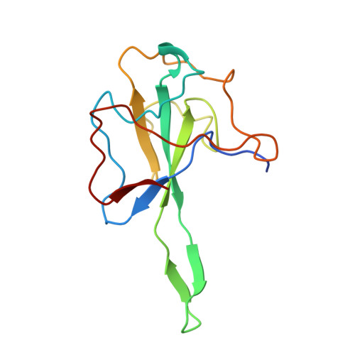
    8X1H

  • PubMed Abstract: 

    The Nucleocapsid (N) protein of SARS-CoV-2 plays a crucial role in viral replication and pathogenesis, making it an attractive target for developing antiviral therapeutics. In this study, we used differential scanning fluorimetry to establish a high-throughput screening method for identifying high-affinity ligands of N-terminal domain of the N protein (N-NTD). We screened an FDA-approved drug library of 1813 compounds and identified 102 compounds interacting with N-NTD. The screened compounds were further investigated for their ability to inhibit the nucleic-acid binding activity of the N protein using electrophoretic mobility-shift assays. We have identified three inhibitors, Ceftazidime, Sennoside A, and Tannic acid, that disrupt the N protein's interaction with RNA probe. Ceftazidime and Sennoside A exhibited nano-molar range binding affinities with N protein, determined through surface plasmon resonance. The binding sites of Ceftazidime and Sennoside A were investigated using [ 1 H, 15 N]-heteronuclear single quantum coherence (HSQC) NMR spectroscopy. Ceftazidime and Sennoside A bind to the putative RNA binding site of the N protein, thus providing insights into the inhibitory mechanism of these compounds. These findings will contribute to the development of novel antiviral agents targeting the N protein of SARS-CoV-2.


  • Organizational Affiliation
    • Protein Crystallography Section, Bhabha Atomic Research Centre, Mumbai, India; Homi Bhabha National Institute, Anushaktinagar, Mumbai, India.

Macromolecule Content 

  • Total Structure Weight: 58.76 kDa 
  • Atom Count: 4,134 
  • Modeled Residue Count: 507 
  • Deposited Residue Count: 536 
  • Unique protein chains: 1

Macromolecules

Find similar proteins by:|  3D Structure
Entity ID: 1
MoleculeChains  Sequence LengthOrganismDetailsImage
Nucleoprotein
A, B, C, D
134Severe acute respiratory syndrome coronavirus 2Mutation(s): 0 
UniProt
Find proteins for P0DTC9 (Severe acute respiratory syndrome coronavirus 2)
Explore P0DTC9 
Go to UniProtKB:  P0DTC9
Entity Groups
Sequence Clusters30% Identity50% Identity70% Identity90% Identity95% Identity100% Identity
UniProt GroupP0DTC9
Sequence Annotations
Expand
Reference Sequence

Experimental Data & Validation

Experimental Data

  • Method: X-RAY DIFFRACTION
  • Resolution: 2.00 Å
  • R-Value Free:  0.278 (Depositor), 0.280 (DCC) 
  • R-Value Work:  0.237 (Depositor), 0.238 (DCC) 
  • R-Value Observed: 0.240 (Depositor) 
Space Group: P 21 21 21
Unit Cell:
Length ( Å )Angle ( ˚ )
a = 58.945α = 90
b = 92.344β = 90
c = 96.014γ = 90
Software Package:
Software NamePurpose
PHENIXrefinement
Aimlessdata scaling
XDSdata reduction
PHENIXphasing

Structure Validation

View Full Validation Report


Entry History 

& Funding Information

Deposition Data


Funding OrganizationLocationGrant Number
Other governmentIndiaNA

Revision History  (Full details and data files)

  • Version 1.0: 2023-11-22
    Type: Initial release
  • Version 1.1: 2024-07-03
    Changes: Database references
  • Version 1.2: 2025-01-15
    Changes: Structure summary
  • Version 2.0: 2025-01-29
    Changes: Atomic model, Data collection, Derived calculations