8WY5

Structure of Gabija GajA in complex with DNA


Experimental Data Snapshot

  • Method: ELECTRON MICROSCOPY
  • Resolution: 3.12 Å
  • Aggregation State: PARTICLE 
  • Reconstruction Method: SINGLE PARTICLE 

wwPDB Validation   3D Report Full Report


This is version 1.1 of the entry. See complete history


Literature

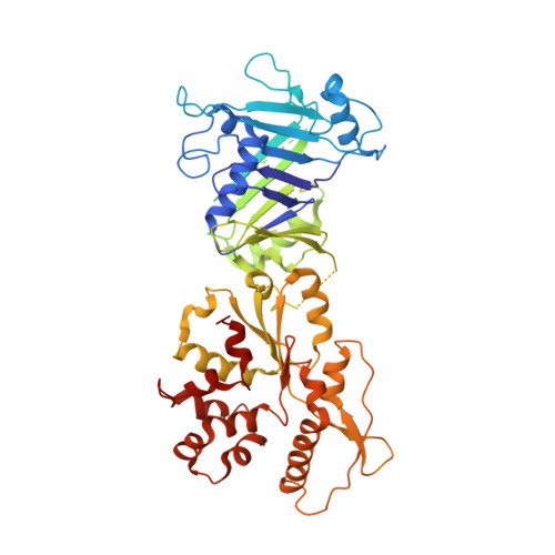
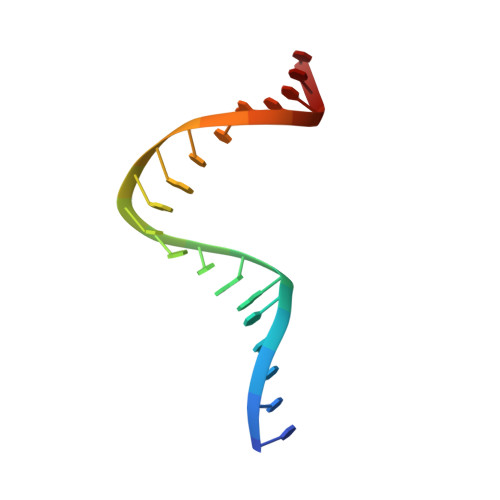
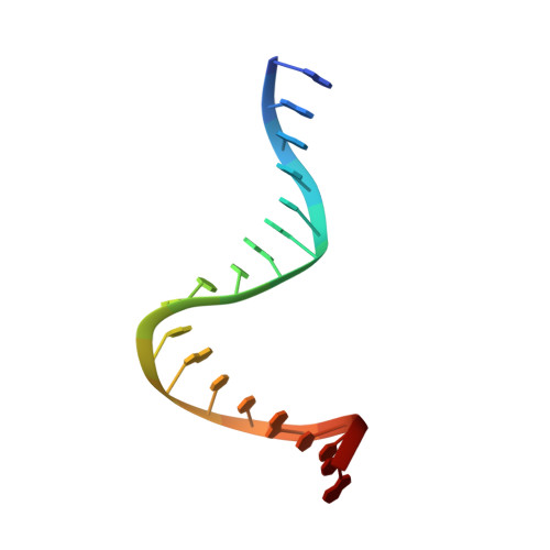
Structures and activation mechanism of the Gabija anti-phage system.

Li, J.Cheng, R.Wang, Z.Yuan, W.Xiao, J.Zhao, X.Du, X.Xia, S.Wang, L.Zhu, B.Wang, L.

(2024) Nature 

  • DOI: https://doi.org/10.1038/s41586-024-07270-x
  • Primary Citation of Related Structures:  
    8JQ9, 8JQB, 8JQC, 8WY4, 8WY5, 8X51, 8X5I, 8X5N

  • PubMed Abstract: 

    Prokaryotes have evolved intricate innate immune systems against phage infection 1-7 . Gabija is a highly widespread prokaryotic defence system that consists of two components, GajA and GajB 8 . GajA functions as a DNA endonuclease that is inactive in the presence of ATP 9 . Here, to explore how the Gabija system is activated for anti-phage defence, we report its cryo-electron microscopy structures in five states, including apo GajA, GajA in complex with DNA, GajA bound by ATP, apo GajA-GajB, and GajA-GajB in complex with ATP and Mg 2+ . GajA is a rhombus-shaped tetramer with its ATPase domain clustered at the centre and the topoisomerase-primase (Toprim) domain located peripherally. ATP binding at the ATPase domain stabilizes the insertion region within the ATPase domain, keeping the Toprim domain in a closed state. Upon ATP depletion by phages, the Toprim domain opens to bind and cleave the DNA substrate. GajB, which docks on GajA, is activated by the cleaved DNA, ultimately leading to prokaryotic cell death. Our study presents a mechanistic landscape of Gabija activation.


  • Organizational Affiliation

    Department of Cardiovascular Surgery, Zhongnan Hospital of Wuhan University, School of Pharmaceutical Sciences, Wuhan University, Wuhan, China.


Macromolecules

Find similar proteins by:  (by identity cutoff)  |  3D Structure
Entity ID: 1
MoleculeChains Sequence LengthOrganismDetailsImage
Endonuclease GajA
A, B, C, D
578Bacillus cereus VD045Mutation(s): 0 
Gene Names: gajAIIE_04982
EC: 3.1
UniProt
Find proteins for J8H9C1 (Bacillus cereus (strain VD045))
Explore J8H9C1 
Go to UniProtKB:  J8H9C1
Entity Groups  
Sequence Clusters30% Identity50% Identity70% Identity90% Identity95% Identity100% Identity
UniProt GroupJ8H9C1
Sequence Annotations
Expand
  • Reference Sequence

Find similar nucleic acids by:  Sequence   |   3D Structure  

Entity ID: 2
MoleculeChains LengthOrganismImage
DNA (5'-D(P*TP*TP*AP*AP*TP*AP*AP*CP*CP*CP*GP*GP*TP*TP*AP*TP*TP*TP*T)-3')
E, G
19synthetic construct
Sequence Annotations
Expand
  • Reference Sequence

Find similar nucleic acids by:  Sequence   |   3D Structure  

Entity ID: 3
MoleculeChains LengthOrganismImage
DNA (5'-D(P*AP*AP*AP*AP*TP*AP*AP*CP*CP*GP*GP*GP*TP*TP*AP*TP*TP*AP*A)-3')
F, H
19synthetic construct
Sequence Annotations
Expand
  • Reference Sequence
Experimental Data & Validation

Experimental Data

  • Method: ELECTRON MICROSCOPY
  • Resolution: 3.12 Å
  • Aggregation State: PARTICLE 
  • Reconstruction Method: SINGLE PARTICLE 
EM Software:
TaskSoftware PackageVersion
MODEL REFINEMENTPHENIX1.20.1_4487:

Structure Validation

View Full Validation Report


Entry History & Funding Information

Deposition Data


Funding OrganizationLocationGrant Number
Not funded--

Revision History  (Full details and data files)

  • Version 1.0: 2024-02-28
    Type: Initial release
  • Version 1.1: 2024-04-03
    Changes: Database references