8WBO | pdb_00008wbo

Crystal structure of cis-Epoxysuccinate Hydrolases RhCESH[L] mutant D18N complexed with sulfate ions


Experimental Data Snapshot

  • Method: X-RAY DIFFRACTION
  • Resolution: 1.58 Å
  • R-Value Free: 
    0.178 (Depositor), 0.180 (DCC) 
  • R-Value Work: 
    0.150 (Depositor), 0.151 (DCC) 
  • R-Value Observed: 
    0.152 (Depositor) 

Starting Model: experimental
View more details

wwPDB Validation 3D Report Full Report

Validation slider image for 8WBO

Ligand Structure Quality Assessment 


This is version 1.2 of the entry. See complete history

Literature

Deciphering the stereo-specific catalytic mechanisms of cis-epoxysuccinate hydrolases producing L(+)-tartaric acid.

Dong, S.Xuan, J.Feng, Y.Cui, Q.

(2024) J Biological Chem 300: 105635-105635

  • DOI: https://doi.org/10.1016/j.jbc.2024.105635
  • Primary Citation Related Structures: 
    8WBK, 8WBL, 8WBM, 8WBN, 8WBO, 8WBP, 8WBQ, 8WBR, 8WBS, 8WBT

  • PubMed Abstract: 

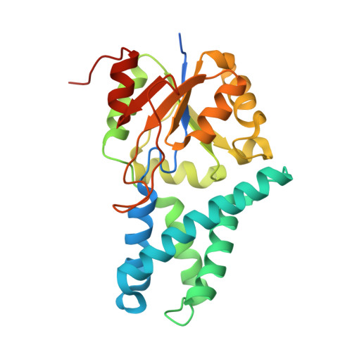
    Microbial epoxide hydrolases, cis-epoxysuccinate hydrolases (CESHs), have been utilized for commercial production of enantiomerically pure L(+)- and D(-)-tartaric acids for decades. However, the stereo-catalytic mechanism of CESH producing L(+)-tartaric acid (CESH[L]) remains unclear. Herein, the crystal structures of two CESH[L]s in ligand-free, product-complexed, and catalytic intermediate forms were determined. These structures revealed the unique specific binding mode for the mirror-symmetric substrate, an active catalytic triad consisting of Asp-His-Glu, and an arginine providing a proton to the oxirane oxygen to facilitate the epoxide ring-opening reaction, which has been pursued for decades. These results provide the structural basis for the rational engineering of these industrial biocatalysts.


  • Organizational Affiliation
    • CAS Key Laboratory of Biofuels, Shandong Provincial Key Laboratory of Synthetic Biology, Qingdao Institute of Bioenergy and Bioprocess Technology, Chinese Academy of Sciences, Qingdao, China; Shandong Energy Institute, Qingdao, China; Qingdao New Energy Shandong Laboratory, Qingdao, China; University of Chinese Academy of Sciences, Beijing, China.

Macromolecule Content 

  • Total Structure Weight: 29.54 kDa 
  • Atom Count: 2,134 
  • Modeled Residue Count: 237 
  • Deposited Residue Count: 264 
  • Unique protein chains: 1

Macromolecules

Find similar proteins by:|  3D Structure
Entity ID: 1
MoleculeChains  Sequence LengthOrganismDetailsImage
Epoxide hydrolase264Rhodococcus opacusMutation(s): 1 
Gene Names: eph
UniProt
Find proteins for Q1KLR5 (Rhodococcus opacus)
Explore Q1KLR5 
Go to UniProtKB:  Q1KLR5
Entity Groups
Sequence Clusters30% Identity50% Identity70% Identity90% Identity95% Identity100% Identity
UniProt GroupQ1KLR5
Sequence Annotations
Expand
Reference Sequence

Experimental Data & Validation

Experimental Data

  • Method: X-RAY DIFFRACTION
  • Resolution: 1.58 Å
  • R-Value Free:  0.178 (Depositor), 0.180 (DCC) 
  • R-Value Work:  0.150 (Depositor), 0.151 (DCC) 
  • R-Value Observed: 0.152 (Depositor) 
Space Group: C 1 2 1
Unit Cell:
Length ( Å )Angle ( ˚ )
a = 63.804α = 90
b = 59.521β = 108.41
c = 68.677γ = 90
Software Package:
Software NamePurpose
HKL-3000data scaling
PHENIXrefinement
HKL-3000data reduction
PHENIXphasing

Structure Validation

View Full Validation Report


Ligand Structure Quality Assessment 


Entry History 

& Funding Information

Deposition Data


Funding OrganizationLocationGrant Number
National Natural Science Foundation of China (NSFC)China32171203

Revision History  (Full details and data files)

  • Version 1.0: 2024-01-31
    Type: Initial release
  • Version 1.1: 2024-02-21
    Changes: Database references
  • Version 1.2: 2024-11-13
    Changes: Structure summary