8WA3

Cryo-EM structure of peptide free and Gs-coupled GIPR


Experimental Data Snapshot

  • Method: ELECTRON MICROSCOPY
  • Resolution: 2.86 Å
  • Aggregation State: PARTICLE 
  • Reconstruction Method: SINGLE PARTICLE 

wwPDB Validation   3D Report Full Report


This is version 1.0 of the entry. See complete history


Literature

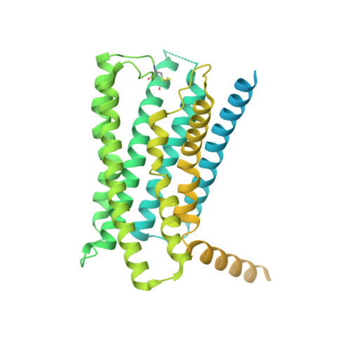
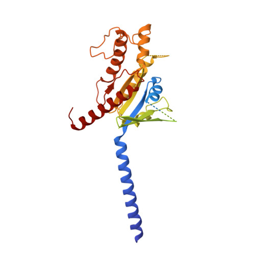
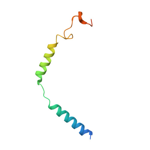
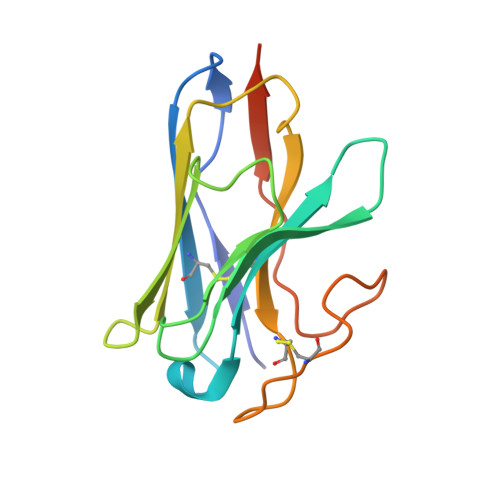
Molecular features of the ligand-free GLP-1R, GCGR and GIPR in complex with G s proteins.

Cong, Z.Zhao, F.Li, Y.Luo, G.Mai, Y.Chen, X.Chen, Y.Lin, S.Cai, X.Zhou, Q.Yang, D.Wang, M.W.

(2024) Cell Discov 10: 18-18

  • DOI: https://doi.org/10.1038/s41421-024-00649-0
  • Primary Citation of Related Structures:  
    8WA3, 8WG7, 8WG8

  • PubMed Abstract: 

    Class B1 G protein-coupled receptors (GPCRs) are important regulators of many physiological functions such as glucose homeostasis, which is mainly mediated by three peptide hormones, i.e., glucagon-like peptide-1 (GLP-1), glucagon (GCG), and glucose-dependent insulinotropic polypeptide (GIP). They trigger a cascade of signaling events leading to the formation of an active agonist-receptor-G protein complex. However, intracellular signal transducers can also activate the receptor independent of extracellular stimuli, suggesting an intrinsic role of G proteins in this process. Here, we report cryo-electron microscopy structures of the human GLP-1 receptor (GLP-1R), GCG receptor (GCGR), and GIP receptor (GIPR) in complex with G s proteins without the presence of cognate ligands. These ligand-free complexes share a similar intracellular architecture to those bound by endogenous peptides, in which, the G s protein alone directly opens the intracellular binding cavity and rewires the extracellular orthosteric pocket to stabilize the receptor in a state unseen before. While the peptide-binding site is partially occupied by the inward folded transmembrane helix 6 (TM6)-extracellular loop 3 (ECL3) juncture of GIPR or a segment of GCGR ECL2, the extracellular portion of GLP-1R adopts a conformation close to the active state. Our findings offer valuable insights into the distinct activation mechanisms of these three important receptors. It is possible that in the absence of a ligand, the intracellular half of transmembrane domain is mobilized with the help of G s protein, which in turn rearranges the extracellular half to form a transitional conformation, facilitating the entry of the peptide N-terminus.


  • Organizational Affiliation

    Department of Pharmacology, School of Basic Medical Sciences, Fudan University, Shanghai, China.


Macromolecules
Find similar proteins by:  (by identity cutoff)  |  3D Structure
Entity ID: 1
MoleculeChains Sequence LengthOrganismDetailsImage
Gastric inhibitory polypeptide receptor,Fusion proteinA [auth R]573Homo sapiensMutation(s): 1 
Gene Names: GIPR
Membrane Entity: Yes 
UniProt & NIH Common Fund Data Resources
Find proteins for P48546 (Homo sapiens)
Explore P48546 
Go to UniProtKB:  P48546
PHAROS:  P48546
GTEx:  ENSG00000010310 
Entity Groups  
Sequence Clusters30% Identity50% Identity70% Identity90% Identity95% Identity100% Identity
UniProt GroupP48546
Sequence Annotations
Expand
  • Reference Sequence
Find similar proteins by:  (by identity cutoff)  |  3D Structure
Entity ID: 2
MoleculeChains Sequence LengthOrganismDetailsImage
Guanine nucleotide-binding protein G(s) subunit alpha isoforms shortB [auth A]394Bos taurusMutation(s): 9 
Gene Names: GNASGNAS1
Membrane Entity: Yes 
UniProt
Find proteins for P04896 (Bos taurus)
Explore P04896 
Go to UniProtKB:  P04896
Entity Groups  
Sequence Clusters30% Identity50% Identity70% Identity90% Identity95% Identity100% Identity
UniProt GroupP04896
Sequence Annotations
Expand
  • Reference Sequence
Find similar proteins by:  (by identity cutoff)  |  3D Structure
Entity ID: 3
MoleculeChains Sequence LengthOrganismDetailsImage
Guanine nucleotide-binding protein G(I)/G(S)/G(T) subunit beta-1,O-antigen polymeraseC [auth B]371Rattus norvegicusLeptolinea tardivitalis
This entity is chimeric
Mutation(s): 0 
Gene Names: Gnb1ADM99_06940
Membrane Entity: Yes 
UniProt
Find proteins for P54311 (Rattus norvegicus)
Explore P54311 
Go to UniProtKB:  P54311
Find proteins for A0A0P6XLS5 (Leptolinea tardivitalis)
Explore A0A0P6XLS5 
Go to UniProtKB:  A0A0P6XLS5
Entity Groups  
Sequence Clusters30% Identity50% Identity70% Identity90% Identity95% Identity100% Identity
UniProt GroupsA0A0P6XLS5P54311
Sequence Annotations
Expand
  • Reference Sequence
Find similar proteins by:  (by identity cutoff)  |  3D Structure
Entity ID: 4
MoleculeChains Sequence LengthOrganismDetailsImage
Guanine nucleotide-binding protein G(I)/G(S)/G(O) subunit gamma-2D [auth G]71Bos taurusMutation(s): 0 
Gene Names: GNG2
Membrane Entity: Yes 
UniProt
Find proteins for P63212 (Bos taurus)
Explore P63212 
Go to UniProtKB:  P63212
Entity Groups  
Sequence Clusters30% Identity50% Identity70% Identity90% Identity95% Identity100% Identity
UniProt GroupP63212
Sequence Annotations
Expand
  • Reference Sequence
Find similar proteins by:  (by identity cutoff)  |  3D Structure
Entity ID: 5
MoleculeChains Sequence LengthOrganismDetailsImage
Nanobody-35E [auth N]140synthetic constructMutation(s): 0 
Entity Groups  
Sequence Clusters30% Identity50% Identity70% Identity90% Identity95% Identity100% Identity
Sequence Annotations
Expand
  • Reference Sequence
Experimental Data & Validation

Experimental Data

  • Method: ELECTRON MICROSCOPY
  • Resolution: 2.86 Å
  • Aggregation State: PARTICLE 
  • Reconstruction Method: SINGLE PARTICLE 

Structure Validation

View Full Validation Report


Entry History & Funding Information

Deposition Data


Funding OrganizationLocationGrant Number
National Natural Science Foundation of China (NSFC)China81872915
National Natural Science Foundation of China (NSFC)China21704064
National Natural Science Foundation of China (NSFC)China81773792
National Natural Science Foundation of China (NSFC)China32071203

Revision History  (Full details and data files)

  • Version 1.0: 2024-03-06
    Type: Initial release