8W31

Crystal structure of parkin (R0RB):2pUb with activator compound


Experimental Data Snapshot

  • Method: X-RAY DIFFRACTION
  • Resolution: 2.50 Å
  • R-Value Free: 0.294 
  • R-Value Work: 0.240 
  • R-Value Observed: 0.242 

Starting Model: experimental
View more details

wwPDB Validation   3D Report Full Report


Ligand Structure Quality Assessment 


This is version 1.1 of the entry. See complete history


Literature

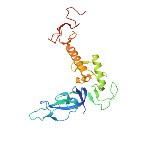
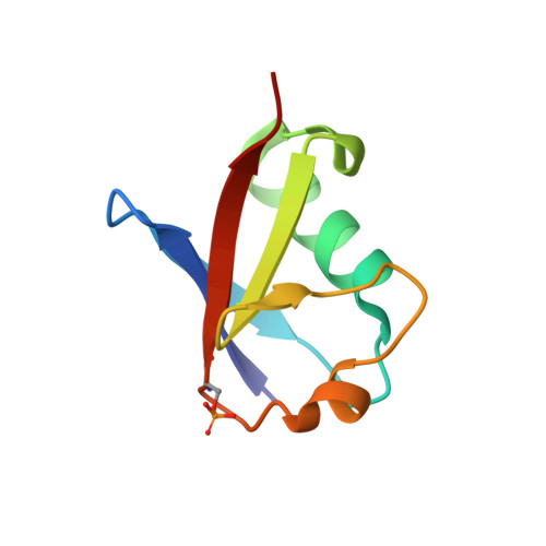
Activation of parkin by a molecular glue.

Sauve, V.Stefan, E.Croteau, N.Goiran, T.Fakih, R.Bansal, N.Hadzipasic, A.Fang, J.Murugan, P.Chen, S.Fon, E.A.Hirst, W.D.Silvian, L.F.Trempe, J.F.Gehring, K.

(2024) Nat Commun 15: 7707-7707

  • DOI: https://doi.org/10.1038/s41467-024-51889-3
  • Primary Citation of Related Structures:  
    8W31

  • PubMed Abstract: 

    Mutations in parkin and PINK1 cause early-onset Parkinson's disease (EOPD). The ubiquitin ligase parkin is recruited to damaged mitochondria and activated by PINK1, a kinase that phosphorylates ubiquitin and the ubiquitin-like domain of parkin. Activated phospho-parkin then ubiquitinates mitochondrial proteins to target the damaged organelle for degradation. Here, we present the mechanism of activation of a new class of small molecule allosteric modulators that enhance parkin activity. The compounds act as molecular glues to enhance the ability of phospho-ubiquitin (pUb) to activate parkin. Ubiquitination assays and isothermal titration calorimetry with the most active compound (BIO-2007817) identify the mechanism of action. We present the crystal structure of a closely related compound (BIO-1975900) bound to a complex of parkin and two pUb molecules. The compound binds next to pUb on RING0 and contacts both proteins. Hydrogen-deuterium exchange mass spectrometry (HDX-MS) experiments confirm that activation occurs through release of the catalytic Rcat domain. In organello and mitophagy assays demonstrate that BIO-2007817 partially rescues the activity of parkin EOPD mutants, R42P and V56E, offering a basis for the design of activators as therapeutics for Parkinson's disease.


  • Organizational Affiliation

    Department of Biochemistry, McGill University, Montreal, QC, Canada.


Macromolecules
Find similar proteins by:  (by identity cutoff)  |  3D Structure
Entity ID: 1
MoleculeChains Sequence LengthOrganismDetailsImage
E3 ubiquitin-protein ligase parkin240Rattus norvegicusMutation(s): 0 
Gene Names: PrknPark2
EC: 2.3.2.31
UniProt
Find proteins for Q9JK66 (Rattus norvegicus)
Explore Q9JK66 
Go to UniProtKB:  Q9JK66
Entity Groups  
Sequence Clusters30% Identity50% Identity70% Identity90% Identity95% Identity100% Identity
UniProt GroupQ9JK66
Sequence Annotations
Expand
  • Reference Sequence
Find similar proteins by:  (by identity cutoff)  |  3D Structure
Entity ID: 2
MoleculeChains Sequence LengthOrganismDetailsImage
Ubiquitin
B, C
75Homo sapiensMutation(s): 0 
Gene Names: UBB
UniProt & NIH Common Fund Data Resources
Find proteins for P0CG47 (Homo sapiens)
Explore P0CG47 
Go to UniProtKB:  P0CG47
PHAROS:  P0CG47
GTEx:  ENSG00000170315 
Entity Groups  
Sequence Clusters30% Identity50% Identity70% Identity90% Identity95% Identity100% Identity
UniProt GroupP0CG47
Sequence Annotations
Expand
  • Reference Sequence
Small Molecules
Ligands 3 Unique
IDChains Name / Formula / InChI Key2D Diagram3D Interactions
A1AE9 (Subject of Investigation/LOI)
Query on A1AE9

Download Ideal Coordinates CCD File 
J [auth A](S)-1-(6-benzyl-3-(4-(1,2,3,4-tetrahydroquinoline-1-carbonyl)phenyl)-6,7-dihydropyrazolo[1,5-a]pyrazin-5(4H)-yl)ethan-1-one
C31 H30 N4 O2
ZYSMPORNWLASGT-MHZLTWQESA-N
GOL
Query on GOL

Download Ideal Coordinates CCD File 
K [auth A]GLYCEROL
C3 H8 O3
PEDCQBHIVMGVHV-UHFFFAOYSA-N
ZN
Query on ZN

Download Ideal Coordinates CCD File 
D [auth A]
E [auth A]
F [auth A]
G [auth A]
H [auth A]
D [auth A],
E [auth A],
F [auth A],
G [auth A],
H [auth A],
I [auth A]
ZINC ION
Zn
PTFCDOFLOPIGGS-UHFFFAOYSA-N
Modified Residues  1 Unique
IDChains TypeFormula2D DiagramParent
SEP
Query on SEP
B, C
L-PEPTIDE LINKINGC3 H8 N O6 PSER
Experimental Data & Validation

Experimental Data

  • Method: X-RAY DIFFRACTION
  • Resolution: 2.50 Å
  • R-Value Free: 0.294 
  • R-Value Work: 0.240 
  • R-Value Observed: 0.242 
  • Space Group: P 61 2 2
Unit Cell:
Length ( Å )Angle ( ˚ )
a = 83.54α = 90
b = 83.54β = 90
c = 258.77γ = 120
Software Package:
Software NamePurpose
PHENIXrefinement
XDSdata scaling
XDSdata reduction
PHENIXphasing
Cootmodel building

Structure Validation

View Full Validation Report


Ligand Structure Quality Assessment 


Entry History & Funding Information

Deposition Data


Funding OrganizationLocationGrant Number
Canadian Institutes of Health Research (CIHR)CanadaFDN 159903

Revision History  (Full details and data files)

  • Version 1.0: 2024-08-21
    Type: Initial release
  • Version 1.1: 2024-10-09
    Changes: Database references, Structure summary