8W26 | pdb_00008w26

X-ray crystal structure of the GAF-PHY domains of SyB-Cph1


Experimental Data Snapshot

  • Method: X-RAY DIFFRACTION
  • Resolution: 2.60 Å
  • R-Value Free: 
    0.242 (Depositor), 0.250 (DCC) 
  • R-Value Work: 
    0.201 (Depositor), 0.200 (DCC) 
  • R-Value Observed: 
    0.205 (Depositor) 

wwPDB Validation   3D Report Full Report


Ligand Structure Quality Assessment 


This is version 1.2 of the entry. See complete history


Literature

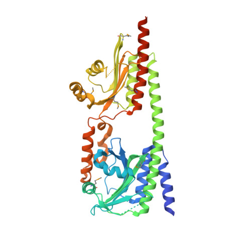
Crystal structure of the photosensory module from a PAS-less cyanobacterial phytochrome as Pr shows a mix of dark-adapted and photoactivated features.

Burgie, E.S.Mickles, A.J.Luo, F.Miller, M.D.Vierstra, R.D.

(2024) J Biological Chem 300: 107369-107369

  • DOI: https://doi.org/10.1016/j.jbc.2024.107369
  • Primary Citation of Related Structures:  
    8W26

  • PubMed Abstract: 

    Phytochromes (Phys) are a diverse collection of photoreceptors that regulate numerous physiological and developmental processes in microorganisms and plants through photointerconversion between red-light-absorbing Pr and far-red light-absorbing Pfr states. Light is detected by an N-terminal photo-sensing module (PSM) sequentially comprised of Period/ARNT/Sim (PAS), cGMP-phosphodiesterase/adenylyl cyclase/FhlA (GAF), and Phy-specific (PHY) domains, with the bilin chromophore covalently-bound within the GAF domain. Phys sense light via the Pr/Pfr ratio measured by the light-induced rotation of the bilin D-pyrrole ring that triggers conformational changes within the PSM, which for microbial Phys reaches into an output region. A key step is a β-stranded to α-helical reconfiguration of a hairpin loop extending from the PHY domain to contact the GAF domain. Besides canonical Phys, cyanobacteria express several variants, including a PAS-less subfamily that harbors just the GAF and PHY domains for light detection. Prior 2D-NMR studies of a model PAS-less Phy from Synechococcus_sp._JA-2-3B'a(2-13) (SyB-Cph1) proposed a unique photoconversion mechanism involving an A-pyrrole ring rotation while magic-angle-spinning NMR probing the chromophore proposed the prototypic D-ring flip. To help solve this conundrum, we determined the crystallographic structure of the GAF-PHY region from SyB-Cph1 as Pr. Surprisingly, this structure differs from canonical Phys by having a Pr ZZZsyn,syn,anti bilin configuration but shifted to the activated position in the binding pocket with consequent folding of the hairpin loop to α-helical, an architecture common for Pfr. Collectively, the PSM of SyB-Cph1 as Pr displayed a mix of dark-adapted and photoactivated features whose co-planar A-C pyrrole rings support a D-ring flip mechanism.


  • Organizational Affiliation
    • Department of Biology, Washington University in St Louis, St Louis, Missouri, USA.

Macromolecules
Find similar proteins by:  (by identity cutoff)  |  3D Structure
Entity ID: 1
MoleculeChains Sequence LengthOrganismDetailsImage
histidine kinase429Synechococcus sp. JA-2-3B'a(2-13)Mutation(s): 0 
Gene Names: CYB_2465
EC: 2.7.13.3
Entity Groups  
Sequence Clusters30% Identity50% Identity70% Identity90% Identity95% Identity100% Identity
Sequence Annotations
Expand
  • Reference Sequence
Small Molecules
Ligands 3 Unique
IDChains Name / Formula / InChI Key2D Diagram3D Interactions
CYC (Subject of Investigation/LOI)
Query on CYC

Download Ideal Coordinates CCD File 
B [auth A]PHYCOCYANOBILIN
C33 H40 N4 O6
VXTXPYZGDQPMHK-GMXXPEQVSA-N
EDO
Query on EDO

Download Ideal Coordinates CCD File 
C [auth A]
D [auth A]
E [auth A]
F [auth A]
G [auth A]
C [auth A],
D [auth A],
E [auth A],
F [auth A],
G [auth A],
H [auth A]
1,2-ETHANEDIOL
C2 H6 O2
LYCAIKOWRPUZTN-UHFFFAOYSA-N
ACT
Query on ACT

Download Ideal Coordinates CCD File 
I [auth A]ACETATE ION
C2 H3 O2
QTBSBXVTEAMEQO-UHFFFAOYSA-M
Modified Residues  1 Unique
IDChains TypeFormula2D DiagramParent
MSE
Query on MSE
A
L-PEPTIDE LINKINGC5 H11 N O2 SeMET
Experimental Data & Validation

Experimental Data

  • Method: X-RAY DIFFRACTION
  • Resolution: 2.60 Å
  • R-Value Free:  0.242 (Depositor), 0.250 (DCC) 
  • R-Value Work:  0.201 (Depositor), 0.200 (DCC) 
  • R-Value Observed: 0.205 (Depositor) 
Space Group: P 43 21 2
Unit Cell:
Length ( Å )Angle ( ˚ )
a = 102.846α = 90
b = 102.846β = 90
c = 130.516γ = 90
Software Package:
Software NamePurpose
PHENIXrefinement
HKL-2000data reduction
HKL-2000data scaling
AutoSolphasing

Structure Validation

View Full Validation Report


Ligand Structure Quality Assessment 


Entry History & Funding Information

Deposition Data


Funding OrganizationLocationGrant Number
National Institutes of Health/National Institute of General Medical Sciences (NIH/NIGMS)United States--

Revision History  (Full details and data files)

  • Version 1.0: 2024-05-22
    Type: Initial release
  • Version 1.1: 2024-10-23
    Changes: Structure summary
  • Version 1.2: 2024-12-25
    Changes: Database references