Regulation of Absorption and Emission in a Protein/Fluorophore Complex.
Santos, E.M., Chandra, I., Assar, Z., Sheng, W., Ghanbarpour, A., Bingham, C., Vasileiou, C., Geiger, J.H., Borhan, B.(2024) ACS Chem Biol 19: 1725-1732
- PubMed: 39046136 Search on PubMed
- DOI: https://doi.org/10.1021/acschembio.4c00125
- Primary Citation Related Structures:
8VZX, 8VZY, 8VZZ, 8W00, 8W02 - PubMed Abstract:
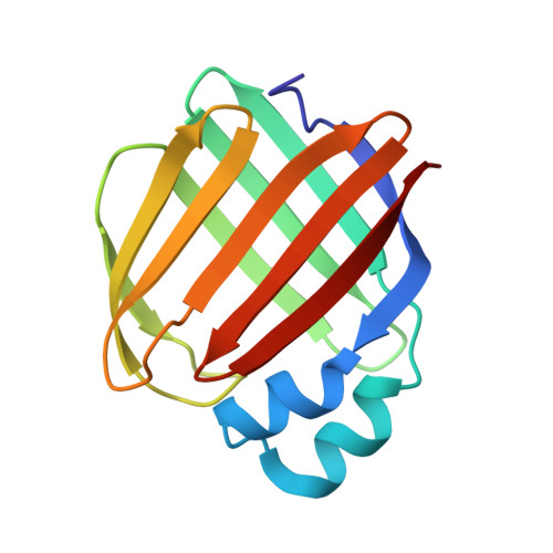
Human cellular retinol binding protein II (hCRBPII) was used as a protein engineering platform to rationally regulate absorptive and emissive properties of a covalently bound fluorogenic dye. We demonstrate the binding of a thio-dapoxyl analog via formation of a protonated imine between an active site lysine residue and the chromophore's aldehyde. Rational manipulation of the electrostatics of the binding pocket results in a 204 nm shift in absorption and a 131 nm shift in emission. The protein is readily expressed in mammalian systems and binds with exogenously delivered fluorophore as demonstrated by live-cell imaging experiments.
- Department of Chemistry, Michigan State University, East Lansing, Michigan 48824, United States.
Organizational Affiliation: