8VWO | pdb_00008vwo

Crystal structure of hRpn13 Pru bound with U-shaped peptide


Experimental Data Snapshot

  • Method: X-RAY DIFFRACTION
  • Resolution: 1.85 Å
  • R-Value Free: 
    0.216 (Depositor), 0.220 (DCC) 
  • R-Value Work: 
    0.195 (Depositor), 0.200 (DCC) 
  • R-Value Observed: 
    0.196 (Depositor) 

Starting Model: experimental
View more details

wwPDB Validation   3D Report Full Report


This is version 1.1 of the entry. See complete history


Literature

An adaptive peptide-binding site in ubiquitin receptor hRpn13 revealed by structural studies.

Hassan, B.Chandravanshi, M.Ng, M.Y.Negi, H.Wilson, B.A.P.Walters, K.J.

(2025) Nat Commun 16: 5669-5669

  • DOI: https://doi.org/10.1038/s41467-025-60843-w
  • Primary Citation of Related Structures:  
    8VWO

  • PubMed Abstract: 

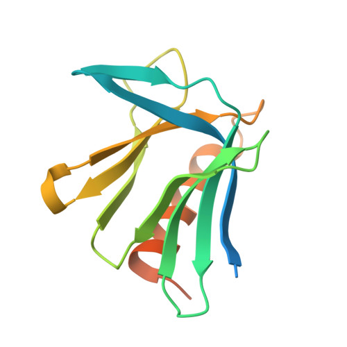
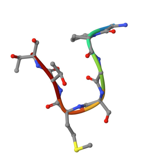
    A pleckstrin-like receptor for ubiquitin (Pru) domain in hRpn13 binds ubiquitin and proteasome subunit hRpn2. Here, we report a crystal structure of Pru bound to amino acids at the extreme N-terminus (ENT) of recombinant hRpn13. ENT adopts a U shape with native sequence along one side where M1 is buried in a Pru W108-centered pocket, and non-native sequence along the other with main chain hydrogen bonding to a neighboring Pru of the crystal lattice. These ENT:Pru interactions are stable in molecular dynamics simulations even with inclusion of only one Pru. Our findings suggest that hRpn13 can form bidentate interactions with ubiquitinated substrates by binding to both ubiquitin chains and disordered sequences of substrates. Testing this model by solution nuclear magnetic resonance revealed Pru to bind weakly to various peptides, concurrent binding with ubiquitin, and ENT displacement by hRpn2, the latter required for substrate handoff to the proteasome ATPases.


  • Organizational Affiliation
    • Protein Processing Section, Center for Structural Biology, Center for Cancer Research, National Cancer Institute, National Institutes of Health, Frederick, MD, USA.

Macromolecules
Find similar proteins by:  (by identity cutoff)  |  3D Structure
Entity ID: 1
MoleculeChains Sequence LengthOrganismDetailsImage
Proteasomal ubiquitin receptor ADRM1150Homo sapiensMutation(s): 0 
Gene Names: ADRM1GP110
UniProt & NIH Common Fund Data Resources
Find proteins for Q16186 (Homo sapiens)
Explore Q16186 
Go to UniProtKB:  Q16186
PHAROS:  Q16186
GTEx:  ENSG00000130706 
Entity Groups  
Sequence Clusters30% Identity50% Identity70% Identity90% Identity95% Identity100% Identity
UniProt GroupQ16186
Sequence Annotations
Expand
  • Reference Sequence

Find similar proteins by:  Sequence   |   3D Structure  

Entity ID: 2
MoleculeChains Sequence LengthOrganismDetailsImage
GLY-PRO-GLY-SER-MET-THR-THR7Homo sapiensMutation(s): 0 
Gene Names: ADRM1
Sequence Annotations
Expand
  • Reference Sequence
Experimental Data & Validation

Experimental Data

  • Method: X-RAY DIFFRACTION
  • Resolution: 1.85 Å
  • R-Value Free:  0.216 (Depositor), 0.220 (DCC) 
  • R-Value Work:  0.195 (Depositor), 0.200 (DCC) 
  • R-Value Observed: 0.196 (Depositor) 
Space Group: P 21 21 21
Unit Cell:
Length ( Å )Angle ( ˚ )
a = 31.262α = 90
b = 56.974β = 90
c = 77.793γ = 90
Software Package:
Software NamePurpose
SERGUIdata collection
DIALSdata reduction
Cootmodel building
REFMACrefinement

Structure Validation

View Full Validation Report


Entry History & Funding Information

Deposition Data


Funding OrganizationLocationGrant Number
National Institutes of Health/National Cancer Institute (NIH/NCI)United States1 ZIABC011490 and FLEX award

Revision History  (Full details and data files)

  • Version 1.0: 2025-07-09
    Type: Initial release
  • Version 1.1: 2025-07-16
    Changes: Database references