Both Domains of APOBEC3F Recognize AA RNA Motifs to Support HIV-1 Virion Encapsidation and Antiviral Function.
Pacheco, J., Yousefi, M., Yang, H., Li, S., Chelico, L., Chen, X.S.(2025) J Mol Biology 438: 169536-169536
- PubMed: 41207371 Search on PubMed
- DOI: https://doi.org/10.1016/j.jmb.2025.169536
- Primary Citation Related Structures:
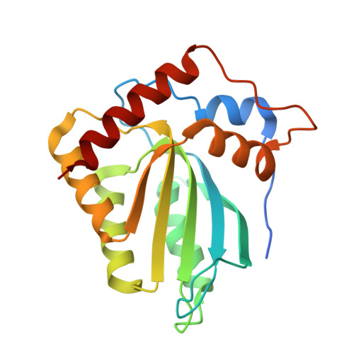
8VUD - PubMed Abstract:
The anti-HIV-1 activity of the double-domain cytidine deaminases APOBEC3G (A3G) and APOBEC3F (A3F) depends on their encapsidation into progeny virions. While A3G requires AA-dinucleotide recognition by its N-terminal deaminase domain (CD1) for packaging, the mechanism for A3F encapsidation has remained unclear. Here, we present the structure of an A3F CD1 variant, revealing AA-binding pocket residues nearly identical to those of A3G CD1. Modeling further shows that A3F's C-terminal deaminase domain (CD2) harbors a similarly conserved AA-binding pocket. Both A3F CD1 and CD2 preferentially bind AA/GA-containing RNA, and mutations in the AA-binding pocket of either domain in full-length A3F do not impair virion packaging or antiviral activity, indicating functional redundancy. Consistently, double-domain chimeras with A3F CD1 or CD2 at either terminus are efficiently packaged and restrict HIV-1 through both deaminase-dependent and -independent mechanisms. In contrast, A3G exhibits strict domain-position dependence: only constructs with A3G CD1 at the N-terminus support packaging, and HIV-restriction activity varies with the particular domain at the C-terminus. A3G CD1 at the C-terminus is inactive, but the A3G CD2 at the C-terminus is active with either the A3F CD1 or A3F CD2 at the N-terminus. These findings highlight the mechanistic flexibility of A3F, in which either domain can mediate RNA recognition, virion encapsidation, and antiviral activity.
- Molecular and Computational Biology, Department of Biological Sciences and Department of Chemistry, University of Southern California, Los Angeles, CA 90089, USA.
Organizational Affiliation: