Identification of FDA-approved drugs that induce heart regeneration in mammals.
Ahmed, M.S., Nguyen, N.U.N., Nakada, Y., Hsu, C.C., Farag, A., Lam, N.T., Wang, P., Thet, S., Menendez-Montes, I., Elhelaly, W.M., Lou, X., Secco, I., Tomczyk, M., Zentilin, L., Pei, J., Cui, M., Dos Santos, M., Liu, X., Liu, Y., Zaha, D., Walcott, G., Tomchick, D.R., Xing, C., Zhang, C.C., Grishin, N.V., Giacca, M., Zhang, J., Sadek, H.A.(2024) Nat Cardiovasc Res 3: 372-388
- PubMed: 39183959 Search on PubMedSearch on PubMed Central
- DOI: https://doi.org/10.1038/s44161-024-00450-y
- Primary Citation Related Structures:
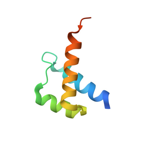
8VTS, 8VTT - PubMed Abstract:
Targeting Meis1 and Hoxb13 transcriptional activity could be a viable therapeutic strategy for heart regeneration. In this study, we performd an in silico screening to identify FDA-approved drugs that can inhibit Meis1 and Hoxb13 transcriptional activity based on the resolved crystal structure of Meis1 and Hoxb13 bound to DNA. Paromomycin (Paro) and neomycin (Neo) induced proliferation of neonatal rat ventricular myocytes in vitro and displayed dose-dependent inhibition of Meis1 and Hoxb13 transcriptional activity by luciferase assay and disruption of DNA binding by electromobility shift assay. X-ray crystal structure revealed that both Paro and Neo bind to Meis1 near the Hoxb13-interacting domain. Administration of Paro-Neo combination in adult mice and in pigs after cardiac ischemia/reperfusion injury induced cardiomyocyte proliferation, improved left ventricular systolic function and decreased scar formation. Collectively, we identified FDA-approved drugs with therapeutic potential for induction of heart regeneration in mammals.
- Division of Cardiology, Department of Internal Medicine, The University of Texas Southwestern Medical Center, Dallas, TX, USA.
Organizational Affiliation: