8VTI | pdb_00008vti

Latrophilin-3 (ADGRL3) HormR and GAIN domains in the context of the holoreceptor


Experimental Data Snapshot

  • Method: ELECTRON MICROSCOPY
  • Resolution: 3.90 Å
  • Aggregation State: PARTICLE 
  • Reconstruction Method: SINGLE PARTICLE 

wwPDB Validation   3D Report Full Report


This is version 1.1 of the entry. See complete history


Literature

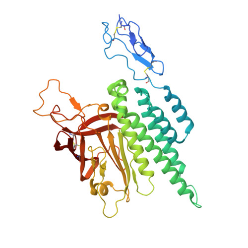
Conformational coupling between extracellular and transmembrane domains modulates holo-adhesion GPCR function.

Kordon, S.P.Cechova, K.Bandekar, S.J.Leon, K.Dutka, P.Siffer, G.Kossiakoff, A.A.Vafabakhsh, R.Arac, D.

(2024) Nat Commun 15: 10545-10545

  • DOI: https://doi.org/10.1038/s41467-024-54836-4
  • Primary Citation of Related Structures:  
    8VTI

  • PubMed Abstract: 

    Adhesion G Protein-Coupled Receptors (aGPCRs) are key cell-adhesion molecules involved in numerous physiological functions. aGPCRs have large multi-domain extracellular regions (ECRs) containing a conserved GAIN domain that precedes their seven-pass transmembrane domain (7TM). Ligand binding and mechanical force applied on the ECR regulate receptor function. However, how the ECR communicates with the 7TM remains elusive, because the relative orientation and dynamics of the ECR and 7TM within a holoreceptor is unclear. Here, we describe the cryo-EM reconstruction of an aGPCR, Latrophilin3/ADGRL3, and reveal that the GAIN domain adopts a parallel orientation to the transmembrane region and has constrained movement. Single-molecule FRET experiments unveil three slow-exchanging FRET states of the ECR relative to the transmembrane region within the holoreceptor. GAIN-targeted antibodies, and cancer-associated mutations at the GAIN-7TM interface, alter FRET states, cryo-EM conformations, and receptor signaling. Altogether, this data demonstrates conformational and functional coupling between the ECR and 7TM, suggesting an ECR-mediated mechanism for aGPCR activation.


  • Organizational Affiliation
    • Department of Biochemistry and Molecular Biology, The University of Chicago, Chicago, IL, USA.

Macromolecules
Find similar proteins by:  (by identity cutoff)  |  3D Structure
Entity ID: 1
MoleculeChains Sequence LengthOrganismDetailsImage
Isoform 4 of Adhesion G protein-coupled receptor L3375Homo sapiensMutation(s): 0 
Gene Names: ADGRL3KIAA0768LEC3LPHN3
UniProt & NIH Common Fund Data Resources
Find proteins for Q9HAR2 (Homo sapiens)
Explore Q9HAR2 
Go to UniProtKB:  Q9HAR2
PHAROS:  Q9HAR2
GTEx:  ENSG00000150471 
Entity Groups  
Sequence Clusters30% Identity50% Identity70% Identity90% Identity95% Identity100% Identity
UniProt GroupQ9HAR2
Glycosylation
Glycosylation Sites: 2Go to GlyGen: Q9HAR2-1
Sequence Annotations
Expand
  • Reference Sequence
Find similar proteins by:  (by identity cutoff)  |  3D Structure
Entity ID: 2
MoleculeChains Sequence LengthOrganismDetailsImage
Isoform 4 of Adhesion G protein-coupled receptor L3314Homo sapiensMutation(s): 0 
Gene Names: ADGRL3KIAA0768LEC3LPHN3
UniProt & NIH Common Fund Data Resources
Find proteins for Q9HAR2 (Homo sapiens)
Explore Q9HAR2 
Go to UniProtKB:  Q9HAR2
PHAROS:  Q9HAR2
GTEx:  ENSG00000150471 
Entity Groups  
Sequence Clusters30% Identity50% Identity70% Identity90% Identity95% Identity100% Identity
UniProt GroupQ9HAR2
Sequence Annotations
Expand
  • Reference Sequence
Find similar proteins by:  (by identity cutoff)  |  3D Structure
Entity ID: 3
MoleculeChains Sequence LengthOrganismDetailsImage
sAB Light Chain215synthetic constructMutation(s): 0 
Entity Groups  
Sequence Clusters30% Identity50% Identity70% Identity90% Identity95% Identity100% Identity
Sequence Annotations
Expand
  • Reference Sequence
Find similar proteins by:  (by identity cutoff)  |  3D Structure
Entity ID: 4
MoleculeChains Sequence LengthOrganismDetailsImage
sAB Heavy Chain235synthetic constructMutation(s): 0 
Entity Groups  
Sequence Clusters30% Identity50% Identity70% Identity90% Identity95% Identity100% Identity
Sequence Annotations
Expand
  • Reference Sequence
Oligosaccharides

Help

Entity ID: 5
MoleculeChains Length2D Diagram Glycosylation3D Interactions
beta-D-mannopyranose-(1-4)-2-acetamido-2-deoxy-beta-D-glucopyranose-(1-4)-2-acetamido-2-deoxy-beta-D-glucopyranose
E
3N-Glycosylation
Glycosylation Resources
GlyTouCan:  G15407YE
GlyCosmos:  G15407YE
GlyGen:  G15407YE
Small Molecules
Ligands 1 Unique
IDChains Name / Formula / InChI Key2D Diagram3D Interactions
NAG
Query on NAG

Download Ideal Coordinates CCD File 
F [auth A]2-acetamido-2-deoxy-beta-D-glucopyranose
C8 H15 N O6
OVRNDRQMDRJTHS-FMDGEEDCSA-N
Experimental Data & Validation

Experimental Data

  • Method: ELECTRON MICROSCOPY
  • Resolution: 3.90 Å
  • Aggregation State: PARTICLE 
  • Reconstruction Method: SINGLE PARTICLE 
EM Software:
TaskSoftware PackageVersion
MODEL REFINEMENTPHENIX

Structure Validation

View Full Validation Report


Entry History & Funding Information

Deposition Data


Funding OrganizationLocationGrant Number
National Institutes of Health/National Institute of General Medical Sciences (NIH/NIGMS)United StatesR35 GM148412
National Institutes of Health/National Institute of General Medical Sciences (NIH/NIGMS)United StatesR01 GM134035-01
National Institutes of Health/National Institute of General Medical Sciences (NIH/NIGMS)United StatesF32 GM142266

Revision History  (Full details and data files)

  • Version 1.0: 2024-12-11
    Type: Initial release
  • Version 1.1: 2025-05-21
    Changes: Data collection