Changes in an enzyme ensemble during catalysis observed by high-resolution XFEL crystallography.
Smith, N., Dasgupta, M., Wych, D.C., Dolamore, C., Sierra, R.G., Lisova, S., Marchany-Rivera, D., Cohen, A.E., Boutet, S., Hunter, M.S., Kupitz, C., Poitevin, F., Moss 3rd, F.R., Mittan-Moreau, D.W., Brewster, A.S., Sauter, N.K., Young, I.D., Wolff, A.M., Tiwari, V.K., Kumar, N., Berkowitz, D.B., Hadt, R.G., Thompson, M.C., Follmer, A.H., Wall, M.E., Wilson, M.A.(2024) Sci Adv 10: eadk7201-eadk7201
- PubMed: 38536910 Search on PubMedSearch on PubMed Central
- DOI: https://doi.org/10.1126/sciadv.adk7201
- Primary Citation Related Structures:
8TSU, 8TSX, 8TSY, 8TSZ, 8TT0, 8TT1, 8TT2, 8TT4, 8TT5, 8VPW, 8VQ1 - PubMed Abstract:
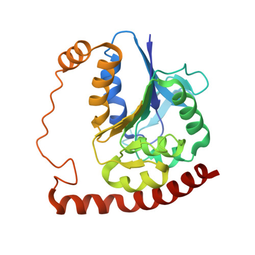
Enzymes populate ensembles of structures necessary for catalysis that are difficult to experimentally characterize. We use time-resolved mix-and-inject serial crystallography at an x-ray free electron laser to observe catalysis in a designed mutant isocyanide hydratase (ICH) enzyme that enhances sampling of important minor conformations. The active site exists in a mixture of conformations, and formation of the thioimidate intermediate selects for catalytically competent substates. The influence of cysteine ionization on the ICH ensemble is validated by determining structures of the enzyme at multiple pH values. Large molecular dynamics simulations in crystallo and time-resolved electron density maps show that Asp 17 ionizes during catalysis and causes conformational changes that propagate across the dimer, permitting water to enter the active site for intermediate hydrolysis. ICH exhibits a tight coupling between ionization of active site residues and catalysis-activated protein motions, exemplifying a mechanism of electrostatic control of enzyme dynamics.
- Department of Biochemistry and Redox Biology Center, University of Nebraska-Lincoln, Lincoln, NE 68588, USA.
Organizational Affiliation: