8VIZ | pdb_00008viz

Structure of full-length gelsolin bound to the barbed end of F-actin


Experimental Data Snapshot

  • Method: ELECTRON MICROSCOPY
  • Resolution: 2.63 Å
  • Aggregation State: PARTICLE 
  • Reconstruction Method: SINGLE PARTICLE 

wwPDB Validation   3D Report Full Report


This is version 1.3 of the entry. See complete history


Literature

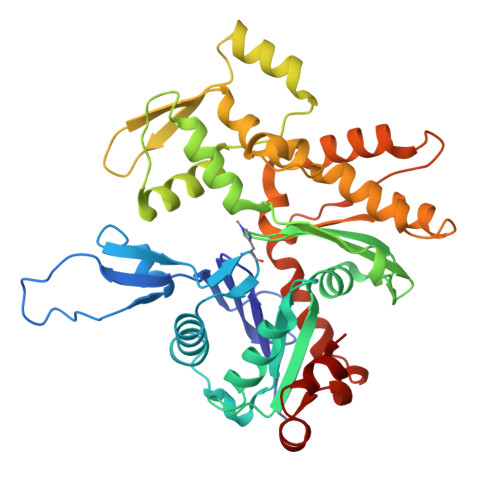
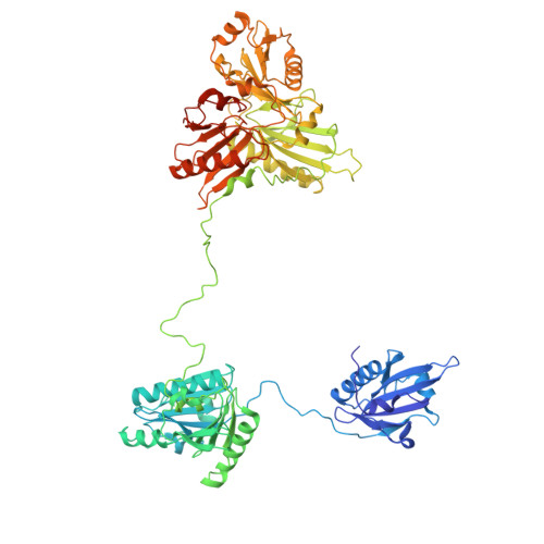
Mechanism of actin filament severing and capping by gelsolin.

Barrie, K.R.Rebowski, G.Dominguez, R.

(2025) Nat Struct Mol Biol 32: 237-242

  • DOI: https://doi.org/10.1038/s41594-024-01412-5
  • Primary Citation of Related Structures:  
    8VIZ, 8VKH

  • PubMed Abstract: 

    Gelsolin is the prototypical member of a family of Ca 2+ -activated F-actin severing and capping proteins. Here we report structures of Ca 2+ -bound human gelsolin at the barbed end of F-actin. One structure reveals gelsolin's six domains (G1G6) and interdomain linkers wrapping around F-actin, while another shows domains G1G3-a fragment observed during apoptosis-binding on both sides of F-actin. Conformational changes that trigger severing occur on one side of F-actin with G1G6 and on both sides with G1G3. Gelsolin remains bound after severing, blocking subunit exchange.


  • Organizational Affiliation
    • Department of Physiology and Biochemistry and Molecular Biophysics Graduate Group, Perelman School of Medicine, University of Pennsylvania, Philadelphia, PA, USA.

Macromolecules
Find similar proteins by:  (by identity cutoff)  |  3D Structure
Entity ID: 1
MoleculeChains Sequence LengthOrganismDetailsImage
Actin, alpha skeletal muscle
A, B, C, D, E
A, B, C, D, E, F
375Oryctolagus cuniculusMutation(s): 0 
EC: 3.6.4
UniProt
Find proteins for P68135 (Oryctolagus cuniculus)
Explore P68135 
Go to UniProtKB:  P68135
Entity Groups  
Sequence Clusters30% Identity50% Identity70% Identity90% Identity95% Identity100% Identity
UniProt GroupP68135
Sequence Annotations
Expand
  • Reference Sequence
Find similar proteins by:  (by identity cutoff)  |  3D Structure
Entity ID: 2
MoleculeChains Sequence LengthOrganismDetailsImage
Gelsolin746Homo sapiensMutation(s): 0 
Gene Names: GSN
UniProt & NIH Common Fund Data Resources
Find proteins for P06396 (Homo sapiens)
Explore P06396 
Go to UniProtKB:  P06396
PHAROS:  P06396
GTEx:  ENSG00000148180 
Entity Groups  
Sequence Clusters30% Identity50% Identity70% Identity90% Identity95% Identity100% Identity
UniProt GroupP06396
Sequence Annotations
Expand
  • Reference Sequence
Small Molecules
Ligands 3 Unique
IDChains Name / Formula / InChI Key2D Diagram3D Interactions
ATP
Query on ATP

Download Ideal Coordinates CCD File 
R [auth F]ADENOSINE-5'-TRIPHOSPHATE
C10 H16 N5 O13 P3
ZKHQWZAMYRWXGA-KQYNXXCUSA-N
ADP
Query on ADP

Download Ideal Coordinates CCD File 
H [auth A],
J [auth B],
L [auth C],
N [auth D],
P [auth E]
ADENOSINE-5'-DIPHOSPHATE
C10 H15 N5 O10 P2
XTWYTFMLZFPYCI-KQYNXXCUSA-N
CA
Query on CA

Download Ideal Coordinates CCD File 
AA [auth G]
I [auth A]
K [auth B]
M [auth C]
O [auth D]
AA [auth G],
I [auth A],
K [auth B],
M [auth C],
O [auth D],
Q [auth E],
S [auth F],
T [auth G],
U [auth G],
V [auth G],
W [auth G],
X [auth G],
Y [auth G],
Z [auth G]
CALCIUM ION
Ca
BHPQYMZQTOCNFJ-UHFFFAOYSA-N
Modified Residues  1 Unique
IDChains TypeFormula2D DiagramParent
HIC
Query on HIC
A, B, C, D, E
A, B, C, D, E, F
L-PEPTIDE LINKINGC7 H11 N3 O2HIS
Experimental Data & Validation

Experimental Data

  • Method: ELECTRON MICROSCOPY
  • Resolution: 2.63 Å
  • Aggregation State: PARTICLE 
  • Reconstruction Method: SINGLE PARTICLE 
EM Software:
TaskSoftware PackageVersion
MODEL REFINEMENTPHENIX

Structure Validation

View Full Validation Report


Entry History & Funding Information

Deposition Data


Funding OrganizationLocationGrant Number
National Institutes of Health/National Institute of General Medical Sciences (NIH/NIGMS)United StatesGM073791

Revision History  (Full details and data files)

  • Version 1.0: 2024-10-16
    Type: Initial release
  • Version 1.1: 2024-11-06
    Changes: Data collection, Database references
  • Version 1.2: 2025-03-05
    Changes: Data collection, Database references
  • Version 1.3: 2025-06-04
    Changes: Data collection