8VHL

Structure of DHODH in Complex with Ligand 17


Experimental Data Snapshot

  • Method: X-RAY DIFFRACTION
  • Resolution: 1.93 Å
  • R-Value Free: 0.199 
  • R-Value Work: 0.168 
  • R-Value Observed: 0.169 

wwPDB Validation   3D Report Full Report


Ligand Structure Quality Assessment 


This is version 1.0 of the entry. See complete history


Literature

Discovery of Alternative Binding Poses through Fragment-Based Identification of DHODH Inhibitors.

DeRatt, L.G.Pietsch, E.C.Cisar, J.S.Jacoby, E.Kazmi, F.Matico, R.Shaffer, P.Tanner, A.Wang, W.Attar, R.Edwards, J.P.Kuduk, S.D.

(2024) ACS Med Chem Lett 15: 381-387

  • DOI: https://doi.org/10.1021/acsmedchemlett.3c00543
  • Primary Citation of Related Structures:  
    8VHL, 8VHM

  • PubMed Abstract: 

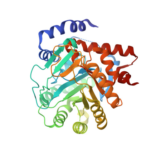
    Dihydroorotate dehydrogenase (DHODH) is a mitochondrial enzyme that affects many aspects essential to cell proliferation and survival. Recently, DHODH has been identified as a potential target for acute myeloid leukemia therapy. Herein, we describe the identification of potent DHODH inhibitors through a scaffold hopping approach emanating from a fragment screen followed by structure-based drug design to further improve the overall profile and reveal an unexpected novel binding mode. Additionally, these compounds had low P-gp efflux ratios, allowing for applications where exposure to the brain would be required.


  • Organizational Affiliation

    Janssen Pharmaceutical Research and Development, 1400 McKean Rd., Spring House, Pennsylvania 19477, United States.


Macromolecules
Find similar proteins by:  (by identity cutoff)  |  3D Structure
Entity ID: 1
MoleculeChains Sequence LengthOrganismDetailsImage
Dihydroorotate dehydrogenase (quinone), mitochondrial369Homo sapiensMutation(s): 0 
Gene Names: DHODH
EC: 1.3.5.2
UniProt & NIH Common Fund Data Resources
Find proteins for Q02127 (Homo sapiens)
Explore Q02127 
Go to UniProtKB:  Q02127
PHAROS:  Q02127
GTEx:  ENSG00000102967 
Entity Groups  
Sequence Clusters30% Identity50% Identity70% Identity90% Identity95% Identity100% Identity
UniProt GroupQ02127
Sequence Annotations
Expand
  • Reference Sequence
Small Molecules
Ligands 6 Unique
IDChains Name / Formula / InChI Key2D Diagram3D Interactions
A1AA2 (Subject of Investigation/LOI)
Query on A1AA2

Download Ideal Coordinates CCD File 
D [auth A]N-(2-chloro-6-fluorophenyl)-4-[ethyl(2-hydroxyethyl)carbamamido]-5-fluoro-2-{[(2S)-1,1,1-trifluoropropan-2-yl]oxy}benzamide
C21 H21 Cl F5 N3 O4
LBIUCQQLEKERKJ-NSHDSACASA-N
FMN
Query on FMN

Download Ideal Coordinates CCD File 
B [auth A]FLAVIN MONONUCLEOTIDE
C17 H21 N4 O9 P
FVTCRASFADXXNN-SCRDCRAPSA-N
ORO
Query on ORO

Download Ideal Coordinates CCD File 
C [auth A]OROTIC ACID
C5 H4 N2 O4
PXQPEWDEAKTCGB-UHFFFAOYSA-N
SO4
Query on SO4

Download Ideal Coordinates CCD File 
G [auth A],
K [auth A],
L [auth A]
SULFATE ION
O4 S
QAOWNCQODCNURD-UHFFFAOYSA-L
GOL
Query on GOL

Download Ideal Coordinates CCD File 
I [auth A],
J [auth A],
M [auth A],
N [auth A]
GLYCEROL
C3 H8 O3
PEDCQBHIVMGVHV-UHFFFAOYSA-N
ACT
Query on ACT

Download Ideal Coordinates CCD File 
E [auth A],
F [auth A],
H [auth A]
ACETATE ION
C2 H3 O2
QTBSBXVTEAMEQO-UHFFFAOYSA-M
Experimental Data & Validation

Experimental Data

  • Method: X-RAY DIFFRACTION
  • Resolution: 1.93 Å
  • R-Value Free: 0.199 
  • R-Value Work: 0.168 
  • R-Value Observed: 0.169 
  • Space Group: P 32 2 1
Unit Cell:
Length ( Å )Angle ( ˚ )
a = 90.744α = 90
b = 90.744β = 90
c = 122.713γ = 120
Software Package:
Software NamePurpose
XDSdata reduction
XSCALEdata scaling
REFMACrefinement
PHASERphasing

Structure Validation

View Full Validation Report


Ligand Structure Quality Assessment 


Entry History & Funding Information

Deposition Data


Funding OrganizationLocationGrant Number
Not funded--

Revision History  (Full details and data files)

  • Version 1.0: 2024-04-03
    Type: Initial release