8V1V | pdb_00008v1v

Human DNA Ligase I F872L bound to adenylated nicked DNA


Experimental Data Snapshot

  • Method: X-RAY DIFFRACTION
  • Resolution: 2.30 Å
  • R-Value Free: 
    0.223 (Depositor), 0.222 (DCC) 
  • R-Value Work: 
    0.179 (Depositor), 0.179 (DCC) 
  • R-Value Observed: 
    0.181 (Depositor) 

Starting Model: experimental
View more details

wwPDB Validation   3D Report Full Report


Ligand Structure Quality Assessment 


This is version 1.1 of the entry. See complete history


Literature

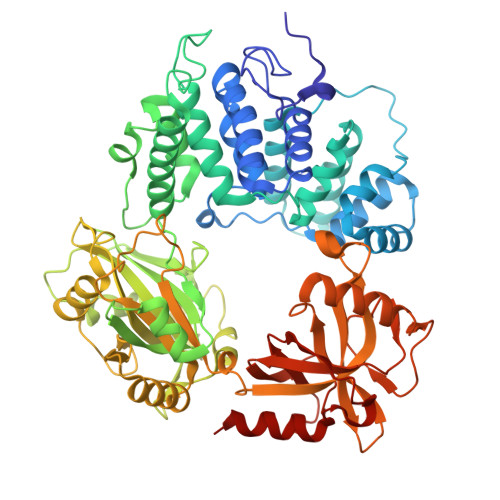
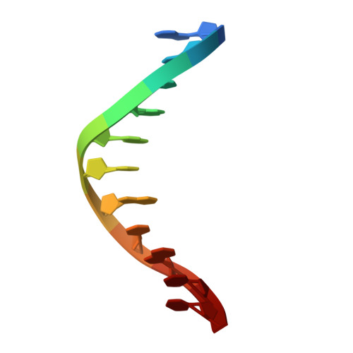
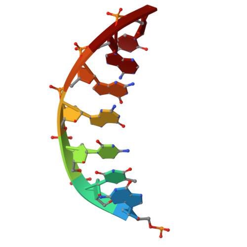
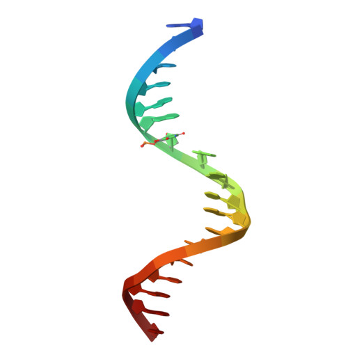
Molecular basis for RNA discrimination by human DNA ligase 1.

Tumbale, P.P.Jurkiw, T.J.Krahn, J.M.Bokil, N.V.Admiraal, S.J.Pedersen, L.C.Williams, J.S.Kunkel, T.A.O'Brien, P.J.Williams, R.S.

(2025) Nucleic Acids Res 53

  • DOI: https://doi.org/10.1093/nar/gkaf299
  • Primary Citation of Related Structures:  
    8V1U, 8V1V, 8V1W

  • PubMed Abstract: 

    DNA ligase 1 (LIG1) finalizes DNA replication and repair by catalyzing the joining of DNA nicks. LIG1 is highly specific for DNA-DNA junctions over DNA-RNA junctions, discriminating strongly against a single ribonucleotide at the 5' side of the nick. This selectivity of LIG1 prevents futile and potentially mutagenic DNA-RNA cleavage and re-ligation cycles during Okazaki fragment maturation or ribonucleotide excision repair of genome-embedded ribonucleotide monophosphates (rNMPs), but the determinants of LIG1 rNMP discrimination are ill-defined. We report structural and kinetic analysis of LIG1 DNA-RNA complexes showing that LIG1 employs an aromatic steric gate to stabilize the enzyme-substrate complex and directly exclude rNMP-containing polynucleotides. Mutation of this RNA gate compromises the adenylyl-transfer and nick-sealing reactions but decreases the discrimination against an rNMP-containing substrate by ∼3600-fold. Our results establish the role of the conserved steric gate in ribonucleotide discrimination by high-fidelity (HiFi) DNA ligases at each step of the ligation reaction, which has parallels to the ribonucleotide discrimination by HiFi DNA polymerases.


  • Organizational Affiliation
    • Genome Integrity and Structural Biology Laboratory, National Institute of Environmental Health Sciences, US National Institutes of Health, Department of Health and Human Services, 111 TW Alexander Drive, Research Triangle Park, NC 27709, United States.

Macromolecules

Find similar proteins by:  (by identity cutoff)  |  3D Structure
Entity ID: 1
MoleculeChains Sequence LengthOrganismDetailsImage
DNA ligase 1647Homo sapiensMutation(s): 1 
Gene Names: LIG1
EC: 6.5.1.1
UniProt & NIH Common Fund Data Resources
Find proteins for P18858 (Homo sapiens)
Explore P18858 
Go to UniProtKB:  P18858
PHAROS:  P18858
GTEx:  ENSG00000105486 
Entity Groups  
Sequence Clusters30% Identity50% Identity70% Identity90% Identity95% Identity100% Identity
UniProt GroupP18858
Sequence Annotations
Expand
  • Reference Sequence

Find similar nucleic acids by:  Sequence   |   3D Structure  

Entity ID: 2
MoleculeChains LengthOrganismImage
DNA (5'-D(*GP*CP*TP*GP*AP*TP*GP*CP*GP*TP*C)-3')11synthetic construct
Sequence Annotations
Expand
  • Reference Sequence

Find similar nucleic acids by:  Sequence   |   3D Structure  

Entity ID: 3
MoleculeChains LengthOrganismImage
DNA (5'-D(P*GP*TP*CP*GP*GP*AP*C)-3')7synthetic construct
Sequence Annotations
Expand
  • Reference Sequence

Find similar nucleic acids by:  Sequence   |   3D Structure  

Entity ID: 4
MoleculeChains LengthOrganismImage
DNA (5'-D(*GP*TP*CP*CP*GP*AP*(OHU)P*GP*AP*CP*GP*CP*AP*TP*CP*AP*GP*C)-3')18synthetic construct
Sequence Annotations
Expand
  • Reference Sequence
Small Molecules
Ligands 4 Unique
IDChains Name / Formula / InChI Key2D Diagram3D Interactions
AMP
Query on AMP

Download Ideal Coordinates CCD File 
I [auth C]ADENOSINE MONOPHOSPHATE
C10 H14 N5 O7 P
UDMBCSSLTHHNCD-KQYNXXCUSA-N
MES (Subject of Investigation/LOI)
Query on MES

Download Ideal Coordinates CCD File 
F [auth A],
K [auth D]
2-(N-MORPHOLINO)-ETHANESULFONIC ACID
C6 H13 N O4 S
SXGZJKUKBWWHRA-UHFFFAOYSA-N
GOL
Query on GOL

Download Ideal Coordinates CCD File 
E [auth A],
G [auth A],
H [auth A]
GLYCEROL
C3 H8 O3
PEDCQBHIVMGVHV-UHFFFAOYSA-N
NA
Query on NA

Download Ideal Coordinates CCD File 
J [auth C]SODIUM ION
Na
FKNQFGJONOIPTF-UHFFFAOYSA-N
Experimental Data & Validation

Experimental Data

  • Method: X-RAY DIFFRACTION
  • Resolution: 2.30 Å
  • R-Value Free:  0.223 (Depositor), 0.222 (DCC) 
  • R-Value Work:  0.179 (Depositor), 0.179 (DCC) 
  • R-Value Observed: 0.181 (Depositor) 
Space Group: P 21 21 21
Unit Cell:
Length ( Å )Angle ( ˚ )
a = 72.966α = 90
b = 96.582β = 90
c = 114.767γ = 90
Software Package:
Software NamePurpose
PHENIXrefinement
PHENIXrefinement
HKL-2000data reduction
HKL-2000data scaling
PHASERphasing

Structure Validation

View Full Validation Report


Ligand Structure Quality Assessment 


Entry History & Funding Information

Deposition Data


Funding OrganizationLocationGrant Number
National Institutes of Health/National Institute of Environmental Health Sciences (NIH/NIEHS)United States1Z01ES102765

Revision History  (Full details and data files)

  • Version 1.0: 2025-07-02
    Type: Initial release
  • Version 1.1: 2026-01-14
    Changes: Database references