8UXS | pdb_00008uxs

KLHDC2 ubiquitin ligase in complex with a novel small-molecule


Experimental Data Snapshot

  • Method: X-RAY DIFFRACTION
  • Resolution: 2.00 Å
  • R-Value Free: 
    0.202 (Depositor), 0.200 (DCC) 
  • R-Value Work: 
    0.166 (Depositor), 0.170 (DCC) 
  • R-Value Observed: 
    0.167 (Depositor) 

Starting Model: experimental
View more details

wwPDB Validation   3D Report Full Report


Ligand Structure Quality Assessment 


This is version 1.1 of the entry. See complete history


Literature

An artificial intelligence accelerated virtual screening platform for drug discovery.

Zhou, G.Rusnac, D.V.Park, H.Canzani, D.Nguyen, H.M.Stewart, L.Bush, M.F.Nguyen, P.T.Wulff, H.Yarov-Yarovoy, V.Zheng, N.DiMaio, F.

(2024) Nat Commun 15: 7761-7761

  • DOI: https://doi.org/10.1038/s41467-024-52061-7
  • Primary Citation of Related Structures:  
    8UXS

  • PubMed Abstract: 

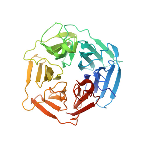
    Structure-based virtual screening is a key tool in early drug discovery, with growing interest in the screening of multi-billion chemical compound libraries. However, the success of virtual screening crucially depends on the accuracy of the binding pose and binding affinity predicted by computational docking. Here we develop a highly accurate structure-based virtual screen method, RosettaVS, for predicting docking poses and binding affinities. Our approach outperforms other state-of-the-art methods on a wide range of benchmarks, partially due to our ability to model receptor flexibility. We incorporate this into a new open-source artificial intelligence accelerated virtual screening platform for drug discovery. Using this platform, we screen multi-billion compound libraries against two unrelated targets, a ubiquitin ligase target KLHDC2 and the human voltage-gated sodium channel Na V 1.7. For both targets, we discover hit compounds, including seven hits (14% hit rate) to KLHDC2 and four hits (44% hit rate) to Na V 1.7, all with single digit micromolar binding affinities. Screening in both cases is completed in less than seven days. Finally, a high resolution X-ray crystallographic structure validates the predicted docking pose for the KLHDC2 ligand complex, demonstrating the effectiveness of our method in lead discovery.


  • Organizational Affiliation
    • Department of Biochemistry, University of Washington, Seattle, WA, USA.

Macromolecules
Find similar proteins by:  (by identity cutoff)  |  3D Structure
Entity ID: 1
MoleculeChains Sequence LengthOrganismDetailsImage
Kelch domain-containing protein 2
A, B
341Homo sapiensMutation(s): 0 
Gene Names: KLHDC2
UniProt & NIH Common Fund Data Resources
Find proteins for Q9Y2U9 (Homo sapiens)
Explore Q9Y2U9 
Go to UniProtKB:  Q9Y2U9
PHAROS:  Q9Y2U9
GTEx:  ENSG00000165516 
Entity Groups  
Sequence Clusters30% Identity50% Identity70% Identity90% Identity95% Identity100% Identity
UniProt GroupQ9Y2U9
Sequence Annotations
Expand
  • Reference Sequence
Small Molecules
Ligands 1 Unique
IDChains Name / Formula / InChI Key2D Diagram3D Interactions
XU8 (Subject of Investigation/LOI)
Query on XU8

Download Ideal Coordinates CCD File 
C [auth A],
D [auth B]
{4-[(2-{[(4-tert-butylphenyl)methyl]sulfanyl}acetamido)methyl]-1H-1,2,3-triazol-1-yl}acetic acid
C18 H24 N4 O3 S
HCTZVVOCYIXIAH-UHFFFAOYSA-N
Experimental Data & Validation

Experimental Data

  • Method: X-RAY DIFFRACTION
  • Resolution: 2.00 Å
  • R-Value Free:  0.202 (Depositor), 0.200 (DCC) 
  • R-Value Work:  0.166 (Depositor), 0.170 (DCC) 
  • R-Value Observed: 0.167 (Depositor) 
Space Group: P 1 21 1
Unit Cell:
Length ( Å )Angle ( ˚ )
a = 44.381α = 90
b = 88.165β = 104.39
c = 88.512γ = 90
Software Package:
Software NamePurpose
PHENIXrefinement
DIALSdata scaling
PHENIXphasing
Cootmodel building

Structure Validation

View Full Validation Report


Ligand Structure Quality Assessment 


Entry History & Funding Information

Deposition Data


Funding OrganizationLocationGrant Number
Howard Hughes Medical Institute (HHMI)United States--

Revision History  (Full details and data files)

  • Version 1.0: 2024-07-10
    Type: Initial release
  • Version 1.1: 2025-01-22
    Changes: Database references, Structure summary