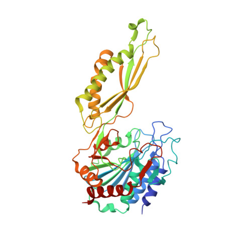
8UW6

Acetylornithine deacetylase from Escherichia coli, di-zinc form.


Experimental Data Snapshot

  • Method: X-RAY DIFFRACTION
  • Resolution: 1.80 Å
  • R-Value Free: 0.203 
  • R-Value Work: 0.163 
  • R-Value Observed: 0.165 

Starting Model: experimental
View more details

wwPDB Validation   3D Report Full Report


This is version 1.1 of the entry. See complete history


Literature

N alpha-acetyl-L-ornithine deacetylase from Escherichia coli and a ninhydrin-based assay to enable inhibitor identification.

Kelley, E.H.Osipiuk, J.Korbas, M.Endres, M.Bland, A.Ehrman, V.Joachimiak, A.Olsen, K.W.Becker, D.P.

(2024) Front Chem 12: 1415644-1415644

  • DOI: https://doi.org/10.3389/fchem.2024.1415644
  • Primary Citation of Related Structures:  
    8UW6

  • PubMed Abstract: 

    Bacteria are becoming increasingly resistant to antibiotics, therefore there is an urgent need for new classes of antibiotics to fight antibiotic resistance. Mammals do not express N ɑ -acetyl-L-ornithine deacetylase (ArgE), an enzyme that is critical for bacterial survival and growth, thus ArgE represents a promising new antibiotic drug target, as inhibitors would not suffer from mechanism-based toxicity. A new ninhydrin-based assay was designed and validated that included the synthesis of the substrate analog N 5 , N 5 -di-methyl N α -acetyl-L-ornithine (k cat /K m = 7.32 ± 0.94 × 10 4  M -1 s -1 ). This new assay enabled the screening of potential inhibitors that absorb in the UV region, and thus is superior to the established 214 nm assay. Using this new ninhydrin-based assay, captopril was confirmed as an ArgE inhibitor (IC 50 = 58.7 μM; K i = 37.1 ± 0.85 μM), and a number of phenylboronic acid derivatives were identified as inhibitors, including 4-(diethylamino)phenylboronic acid (IC 50 = 50.1 μM). Selected inhibitors were also tested in a thermal shift assay with ArgE using SYPRO Orange dye against Escherichia coli ArgE to observe the stability of the enzyme in the presence of inhibitors (captopril K i = 35.9 ± 5.1 μM). The active site structure of di-Zn Ec ArgE was confirmed using X-ray absorption spectroscopy, and we reported two X-ray crystal structures of E. coli ArgE. In summary, we describe the development of a new ninhydrin-based assay for ArgE, the identification of captopril and phenylboronic acids as ArgE inhibitors, thermal shift studies with ArgE + captopril, and the first two published crystal structures of ArgE (mono-Zn and di-Zn).


  • Organizational Affiliation

    Department of Chemistry and Biochemistry, Loyola University Chicago, Chicago, IL, United States.


Macromolecules
Find similar proteins by:  (by identity cutoff)  |  3D Structure
Entity ID: 1
MoleculeChains Sequence LengthOrganismDetailsImage
Acetylornithine deacetylase
A, B, C, D
386Escherichia coli str. K-12 substr. MG1655Mutation(s): 0 
Gene Names: argEZ5515ECs4886
EC: 3.5.1.16
UniProt
Find proteins for P23908 (Escherichia coli (strain K12))
Explore P23908 
Go to UniProtKB:  P23908
Entity Groups  
Sequence Clusters30% Identity50% Identity70% Identity90% Identity95% Identity100% Identity
UniProt GroupP23908
Sequence Annotations
Expand
  • Reference Sequence
Small Molecules
Ligands 4 Unique
IDChains Name / Formula / InChI Key2D Diagram3D Interactions
TRS
Query on TRS

Download Ideal Coordinates CCD File 
AA [auth D]
I [auth A]
O [auth B]
P [auth B]
T [auth C]
AA [auth D],
I [auth A],
O [auth B],
P [auth B],
T [auth C],
Z [auth D]
2-AMINO-2-HYDROXYMETHYL-PROPANE-1,3-DIOL
C4 H12 N O3
LENZDBCJOHFCAS-UHFFFAOYSA-O
SO4
Query on SO4

Download Ideal Coordinates CCD File 
G [auth A],
H [auth A],
K [auth A],
S [auth C],
V [auth C]
SULFATE ION
O4 S
QAOWNCQODCNURD-UHFFFAOYSA-L
ZN (Subject of Investigation/LOI)
Query on ZN

Download Ideal Coordinates CCD File 
E [auth A]
F [auth A]
L [auth B]
M [auth B]
N [auth B]
E [auth A],
F [auth A],
L [auth B],
M [auth B],
N [auth B],
Q [auth C],
R [auth C],
W [auth D],
X [auth D],
Y [auth D]
ZINC ION
Zn
PTFCDOFLOPIGGS-UHFFFAOYSA-N
EDO
Query on EDO

Download Ideal Coordinates CCD File 
J [auth A],
U [auth C]
1,2-ETHANEDIOL
C2 H6 O2
LYCAIKOWRPUZTN-UHFFFAOYSA-N
Experimental Data & Validation

Experimental Data

  • Method: X-RAY DIFFRACTION
  • Resolution: 1.80 Å
  • R-Value Free: 0.203 
  • R-Value Work: 0.163 
  • R-Value Observed: 0.165 
  • Space Group: P 1 21 1
Unit Cell:
Length ( Å )Angle ( ˚ )
a = 52.399α = 90
b = 126.323β = 90.88
c = 123.408γ = 90
Software Package:
Software NamePurpose
REFMACrefinement
HKL-3000data scaling
HKL-3000data reduction
HKL-3000phasing

Structure Validation

View Full Validation Report


Entry History & Funding Information

Deposition Data


Funding OrganizationLocationGrant Number
National Institutes of Health/National Institute Of Allergy and Infectious Diseases (NIH/NIAID)United States75N93022C00035

Revision History  (Full details and data files)

  • Version 1.0: 2024-05-29
    Type: Initial release
  • Version 1.1: 2024-08-07
    Changes: Database references