


X-ray-driven chemistry and conformational heterogeneity in atomic resolution crystal structures of bacterial dihydrofolate reductases
Smith, N., Horswill, A.R., Wilson, M.A.To be published.
Experimental Data Snapshot
Starting Model: experimental
View more details
Entity ID: 1 | |||||
|---|---|---|---|---|---|
| Molecule | Chains | Sequence Length | Organism | Details | Image |
| Dihydrofolate reductase | 159 | Escherichia coli | Mutation(s): 0 Gene Names: folA EC: 1.5.1.3 | 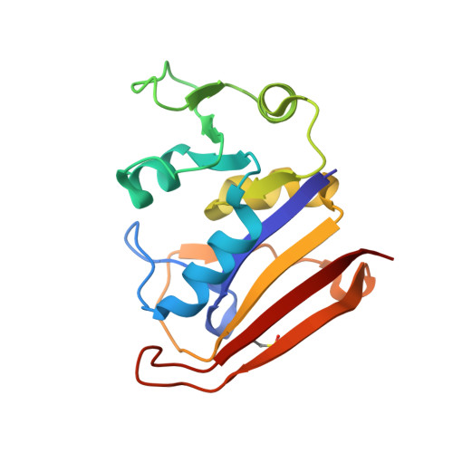 | |
UniProt | |||||
Entity Groups | |||||
| Sequence Clusters | 30% Identity50% Identity70% Identity90% Identity95% Identity100% Identity | ||||
| UniProt Group | P0ABQ4 | ||||
Sequence AnnotationsExpand | |||||
Reference Sequence | |||||
| Ligands 3 Unique | |||||
|---|---|---|---|---|---|
| ID | Chains | Name / Formula / InChI Key | 2D Diagram | 3D Interactions | |
| NAP (Subject of Investigation/LOI) Download:Ideal Coordinates CCD File | C [auth A] | NADP NICOTINAMIDE-ADENINE-DINUCLEOTIDE PHOSPHATE C21 H28 N7 O17 P3 XJLXINKUBYWONI-NNYOXOHSSA-N |  | ||
| FOL (Subject of Investigation/LOI) Download:Ideal Coordinates CCD File | B [auth A] | FOLIC ACID C19 H19 N7 O6 OVBPIULPVIDEAO-LBPRGKRZSA-N |  | ||
| MN Download:Ideal Coordinates CCD File | D [auth A], E [auth A], F [auth A], G [auth A] | MANGANESE (II) ION Mn WAEMQWOKJMHJLA-UHFFFAOYSA-N |  | ||
| Modified Residues 1 Unique | |||||
|---|---|---|---|---|---|
| ID | Chains | Type | Formula | 2D Diagram | Parent |
| OCS Query on OCS | A | L-PEPTIDE LINKING | C3 H7 N O5 S |  | CYS |
| Length ( Å ) | Angle ( ˚ ) |
|---|---|
| a = 33.98 | α = 90 |
| b = 44.848 | β = 90 |
| c = 98.24 | γ = 90 |
| Software Name | Purpose |
|---|---|
| PHENIX | refinement |
| HKL-2000 | data reduction |
| HKL-2000 | data scaling |
| PHENIX | phasing |
| Funding Organization | Location | Grant Number |
|---|---|---|
| National Institutes of Health/National Institute of General Medical Sciences (NIH/NIGMS) | United States | R01GM139978 |


