The reversible activation of norovirus by metal ions.
Sherman, M., Cox, F., Smith, H., Habib, M.H., Karst, S., Wobus, C.E., Smith, T.J.(2024) J Virol 98: e0173523-e0173523
- PubMed: 38236007 Search on PubMedSearch on PubMed Central
- DOI: https://doi.org/10.1128/jvi.01735-23
- Primary Citation Related Structures:
8UUX, 8UV3 - PubMed Abstract:
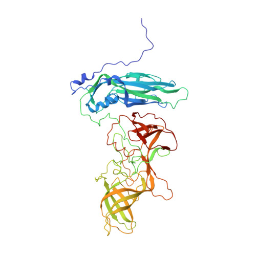
Murine norovirus (MNV) undergoes extremely large conformational changes in response to the environment. The T = 3 icosahedral capsid is composed of 180 copies of ~58-kDa VP1 comprised of N-terminus (N), shell (S), and C-terminal protruding (P) domains. At neutral pH, the P domains are loosely tethered to the shell and float ~15 Å above the surface. At low pH or in the presence of bile salts, the P domain drops onto the shell and this movement is accompanied by conformational changes within the P domain that enhance receptor interactions while blocking antibody binding. While previous crystallographic studies identified metal binding sites in the isolated P domain, the ~2.7-Å cryo-electron microscopy structures of MNV in the presence of Mg 2+ or Ca 2+ presented here show that metal ions can recapitulate the contraction observed at low pH or in the presence of bile. Further, we show that these conformational changes are reversed by dialysis against EDTA. As observed in the P domain crystal structures, metal ions bind to and contract the G'H' loop. This movement is correlated with the lifting of the C'D' loop and rotation of the P domain dimers about each other, exposing the bile salt binding pocket. Isothermal titration calorimetry experiments presented here demonstrate that the activation signals (bile salts, low pH, and metal ions) act in a synergistic manner that, individually, all result in the same activated structure. We present a model whereby these reversible conformational changes represent a uniquely dynamic and tissue-specific structural adaptation to the in vivo environment.IMPORTANCEThe highly mobile protruding domains on the calicivirus capsids are recognized by cell receptor(s) and antibodies. At neutral pH, they float ~15 Å above the shell but at low pH or in the presence of bile salts, they contract onto the surface. Concomitantly, changes within the P domain block antibody binding while enhancing receptor binding. While we previously demonstrated that metals also block antibody binding, it was unknown whether they might also cause similar conformational changes in the virion. Here, we present the near atomic cryo-electron microscopy structures of infectious murine norovirus (MNV) in the presence of calcium or magnesium ions. The metal ions reversibly induce the same P domain contraction as low pH and bile salts and act in a synergistic manner with the other stimuli. We propose that, unlike most other viruses, MNV facilely changes conformations as a unique means to escape immune surveillance as it moves through various tissues.
- Department of Biochemistry and Molecular Biology, University of Texas Medical Branch at Galveston, Galveston, Texas, USA.
Organizational Affiliation: