8UQO

PLCb3-Gbg-Gaq complex on membranes

  • Classification: SIGNALING PROTEIN
  • Organism(s): Homo sapiens
  • Expression System: Trichoplusia ni
  • Mutation(s): No 

  • Deposited: 2023-10-24 Released: 2023-12-06 
  • Deposition Author(s): Falzone, M.E., MacKinnon, R.
  • Funding Organization(s): Howard Hughes Medical Institute (HHMI), National Institutes of Health/National Institute of General Medical Sciences (NIH/NIGMS)

Experimental Data Snapshot

  • Method: ELECTRON MICROSCOPY
  • Resolution: 3.37 Å
  • Aggregation State: PARTICLE 
  • Reconstruction Method: SINGLE PARTICLE 

wwPDB Validation   3D Report Full Report


This is version 1.0 of the entry. See complete history


Literature

The mechanism of G alpha q regulation of PLC beta 3 -catalyzed PIP2 hydrolysis.

Falzone, M.E.MacKinnon, R.

(2023) Proc Natl Acad Sci U S A 120: e2315011120-e2315011120

  • DOI: https://doi.org/10.1073/pnas.2315011120
  • Primary Citation of Related Structures:  
    8UQN, 8UQO

  • PubMed Abstract: 

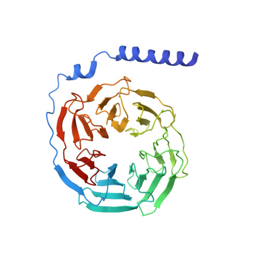
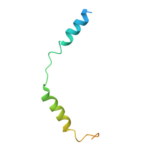
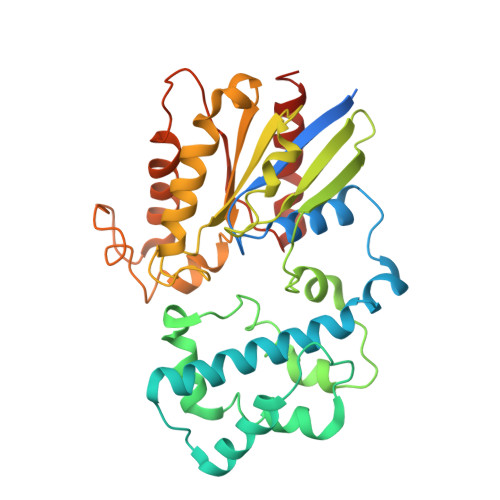
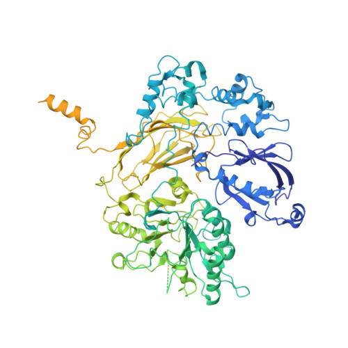
    PLCβ ( Phospholipase Cβ ) enzymes cleave phosphatidylinositol 4,5-bisphosphate ( PIP2) producing IP3 and DAG (diacylglycerol). PIP2 modulates the function of many ion channels, while IP3 and DAG regulate intracellular Ca 2+ levels and protein phosphorylation by protein kinase C, respectively. PLCβ enzymes are under the control of G protein coupled receptor signaling through direct interactions with G proteins Gβγ and q and have been shown to be coincidence detectors for dual stimulation of q and i -coupled receptors. PLCβs are aqueous-soluble cytoplasmic enzymes but partition onto the membrane surface to access their lipid substrate, complicating their functional and structural characterization. Using newly developed methods, we recently showed that Gβγ activates PLCβ3 by recruiting it to the membrane. Using these same methods, here we show that q increases the catalytic rate constant, k cat , of PLCβ3 . Since stimulation of PLCβ3 by q depends on an autoinhibitory element (the X-Y linker), we propose that q produces partial relief of the X-Y linker autoinhibition through an allosteric mechanism. We also determined membrane-bound structures of the PLCβ3·Gα q and PLCβ3·Gβγ (2) ·Gα q complexes, which show that these G proteins can bind simultaneously and independently of each other to regulate PLCβ3 activity. The structures rationalize a finding in the enzyme assay, that costimulation by both G proteins follows a product rule of each independent stimulus. We conclude that baseline activity of PLCβ3 is strongly suppressed, but the effect of G proteins, especially acting together, provides a robust stimulus upon G protein stimulation.


  • Organizational Affiliation

    Laboratory of Molecular Neurobiology and Biophysics, The Rockefeller University, New York, NY 10065.


Macromolecules
Find similar proteins by:  (by identity cutoff)  |  3D Structure
Entity ID: 1
MoleculeChains Sequence LengthOrganismDetailsImage
Guanine nucleotide-binding protein G(I)/G(S)/G(T) subunit beta-1A [auth B],
B [auth C]
340Homo sapiensMutation(s): 0 
Gene Names: GNB1
UniProt & NIH Common Fund Data Resources
Find proteins for P62873 (Homo sapiens)
Explore P62873 
Go to UniProtKB:  P62873
PHAROS:  P62873
GTEx:  ENSG00000078369 
Entity Groups  
Sequence Clusters30% Identity50% Identity70% Identity90% Identity95% Identity100% Identity
UniProt GroupP62873
Sequence Annotations
Expand
  • Reference Sequence
Find similar proteins by:  (by identity cutoff)  |  3D Structure
Entity ID: 2
MoleculeChains Sequence LengthOrganismDetailsImage
Guanine nucleotide-binding protein subunit gammaC [auth D],
D [auth G]
73Homo sapiensMutation(s): 0 
Gene Names: GNG2
UniProt & NIH Common Fund Data Resources
Find proteins for P59768 (Homo sapiens)
Explore P59768 
Go to UniProtKB:  P59768
PHAROS:  P59768
GTEx:  ENSG00000186469 
Entity Groups  
Sequence Clusters30% Identity50% Identity70% Identity90% Identity95% Identity100% Identity
UniProt GroupP59768
Sequence Annotations
Expand
  • Reference Sequence
Find similar proteins by:  (by identity cutoff)  |  3D Structure
Entity ID: 3
MoleculeChains Sequence LengthOrganismDetailsImage
Guanine nucleotide-binding protein G(q) subunit alphaE [auth A]355Homo sapiensMutation(s): 0 
Gene Names: GNAQ
UniProt & NIH Common Fund Data Resources
Find proteins for P50148 (Homo sapiens)
Explore P50148 
Go to UniProtKB:  P50148
PHAROS:  P50148
GTEx:  ENSG00000156052 
Entity Groups  
Sequence Clusters30% Identity50% Identity70% Identity90% Identity95% Identity100% Identity
UniProt GroupP50148
Sequence Annotations
Expand
  • Reference Sequence
Find similar proteins by:  (by identity cutoff)  |  3D Structure
Entity ID: 4
MoleculeChains Sequence LengthOrganismDetailsImage
1-phosphatidylinositol 4,5-bisphosphate phosphodiesterase beta-3F [auth Q]1,234Homo sapiensMutation(s): 0 
Gene Names: PLCB3
EC: 3.1.4.11
UniProt & NIH Common Fund Data Resources
Find proteins for Q01970 (Homo sapiens)
Explore Q01970 
Go to UniProtKB:  Q01970
PHAROS:  Q01970
GTEx:  ENSG00000149782 
Entity Groups  
Sequence Clusters30% Identity50% Identity70% Identity90% Identity95% Identity100% Identity
UniProt GroupQ01970
Sequence Annotations
Expand
  • Reference Sequence
Experimental Data & Validation

Experimental Data

  • Method: ELECTRON MICROSCOPY
  • Resolution: 3.37 Å
  • Aggregation State: PARTICLE 
  • Reconstruction Method: SINGLE PARTICLE 

Structure Validation

View Full Validation Report


Entry History & Funding Information

Deposition Data


Funding OrganizationLocationGrant Number
Howard Hughes Medical Institute (HHMI)United States--
National Institutes of Health/National Institute of General Medical Sciences (NIH/NIGMS)United StatesGM142137

Revision History  (Full details and data files)

  • Version 1.0: 2023-12-06
    Type: Initial release