8ULG | pdb_00008ulg

Cryo-EM structure of bovine phosphodiesterase 6 bound to IBMX


Experimental Data Snapshot

  • Method: ELECTRON MICROSCOPY
  • Resolution: 3.20 Å
  • Aggregation State: PARTICLE 
  • Reconstruction Method: SINGLE PARTICLE 

wwPDB Validation 3D Report Full Report

Validation slider image for 8ULG

This is version 1.1 of the entry. See complete history

Literature

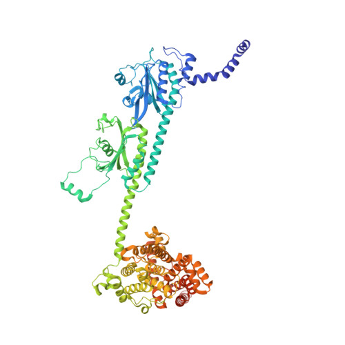
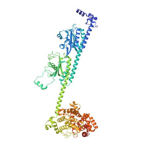
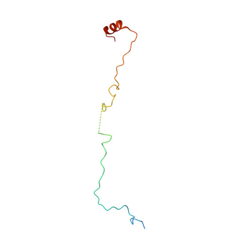
Probing the mechanism by which the retinal G protein transducin activates its biological effector PDE6.

Aplin, C.Cerione, R.A.

(2023) J Biological Chem 300: 105608-105608

  • DOI: https://doi.org/10.1016/j.jbc.2023.105608
  • Primary Citation Related Structures: 
    8UFI, 8UGB, 8UGS, 8ULG

  • PubMed Abstract: 

    Phototransduction in retinal rods occurs when the G protein-coupled photoreceptor rhodopsin triggers the activation of phosphodiesterase 6 (PDE6) by GTP-bound alpha subunits of the G protein transducin (Gα T ). Recently, we presented a cryo-EM structure for a complex between two GTP-bound recombinant Gα T subunits and native PDE6, that included a bivalent antibody bound to the C-terminal ends of Gα T and the inhibitor vardenafil occupying the active sites on the PDEα and PDEβ subunits. We proposed Gα T -activated PDE6 by inducing a striking reorientation of the PDEγ subunits away from the catalytic sites. However, questions remained including whether in the absence of the antibody Gα T binds to PDE6 in a similar manner as observed when the antibody is present, does Gα T activate PDE6 by enabling the substrate cGMP to access the catalytic sites, and how does the lipid membrane enhance PDE6 activation? Here, we demonstrate that 2:1 Gα T -PDE6 complexes form with either recombinant or retinal Gα T in the absence of the Gα T antibody. We show that Gα T binding is not necessary for cGMP nor competitive inhibitors to access the active sites; instead, occupancy of the substrate binding sites enables Gα T to bind and reposition the PDE6γ subunits to promote catalytic activity. Moreover, we demonstrate by reconstituting Gα T -stimulated PDE6 activity in lipid bilayer nanodiscs that the membrane-induced enhancement results from an increase in the apparent binding affinity of Gα T for PDE6. These findings provide new insights into how the retinal G protein stimulates rapid catalytic turnover by PDE6 required for dim light vision.


  • Organizational Affiliation
    • Department of Chemistry and Chemical Biology, Cornell University, Ithaca, New York, USA.

Macromolecule Content 

  • Total Structure Weight: 218.59 kDa 
  • Atom Count: 14,635 
  • Modeled Residue Count: 1,789 
  • Deposited Residue Count: 1,886 
  • Unique protein chains: 3

Macromolecules

Find similar proteins by:|  3D Structure
Entity ID: 1
MoleculeChains  Sequence LengthOrganismDetailsImage
Rod cGMP-specific 3',5'-cyclic phosphodiesterase subunit alpha859Bos taurusMutation(s): 0 
EC: 3.1.4.35
UniProt
Find proteins for P11541 (Bos taurus)
Explore P11541 
Go to UniProtKB:  P11541
Entity Groups
Sequence Clusters30% Identity50% Identity70% Identity90% Identity95% Identity100% Identity
UniProt GroupP11541
Sequence Annotations
Expand
Reference Sequence
Find similar proteins by:|  3D Structure
Entity ID: 2
MoleculeChains  Sequence LengthOrganismDetailsImage
Rod cGMP-specific 3',5'-cyclic phosphodiesterase subunit beta853Bos taurusMutation(s): 0 
EC: 3.1.4.35
UniProt
Find proteins for P23439 (Bos taurus)
Explore P23439 
Go to UniProtKB:  P23439
Entity Groups
Sequence Clusters30% Identity50% Identity70% Identity90% Identity95% Identity100% Identity
UniProt GroupP23439
Sequence Annotations
Expand
Reference Sequence
Find similar proteins by:|  3D Structure
Entity ID: 3
MoleculeChains  Sequence LengthOrganismDetailsImage
Retinal rod rhodopsin-sensitive cGMP 3',5'-cyclic phosphodiesterase subunit gamma
C, D
87Bos taurusMutation(s): 0 
EC: 3.1.4.35
UniProt
Find proteins for P04972 (Bos taurus)
Explore P04972 
Go to UniProtKB:  P04972
Entity Groups
Sequence Clusters30% Identity50% Identity70% Identity90% Identity95% Identity100% Identity
UniProt GroupP04972
Sequence Annotations
Expand
Reference Sequence

Small Molecules

Ligands 4 Unique
IDChains Name / Formula / InChI Key2D Diagram3D Interactions
PCG
(Subject of Investigation/LOI)

Query on PCG


Download:Ideal Coordinates CCD File
E [auth A],
I [auth B]
CYCLIC GUANOSINE MONOPHOSPHATE
C10 H12 N5 O7 P
ZOOGRGPOEVQQDX-UUOKFMHZSA-N
IBM
(Subject of Investigation/LOI)

Query on IBM


Download:Ideal Coordinates CCD File
H [auth A],
L [auth B]
3-ISOBUTYL-1-METHYLXANTHINE
C10 H14 N4 O2
APIXJSLKIYYUKG-UHFFFAOYSA-N
ZN

Query on ZN


Download:Ideal Coordinates CCD File
G [auth A],
K [auth B]
ZINC ION
Zn
PTFCDOFLOPIGGS-UHFFFAOYSA-N
MG

Query on MG


Download:Ideal Coordinates CCD File
F [auth A],
J [auth B]
MAGNESIUM ION
Mg
JLVVSXFLKOJNIY-UHFFFAOYSA-N

Experimental Data & Validation

Experimental Data

  • Method: ELECTRON MICROSCOPY
  • Resolution: 3.20 Å
  • Aggregation State: PARTICLE 
  • Reconstruction Method: SINGLE PARTICLE 

Structure Validation

View Full Validation Report


Entry History 

& Funding Information

Deposition Data


Funding OrganizationLocationGrant Number
National Institutes of Health/National Eye Institute (NIH/NEI)United States1R01EY034867-01

Revision History  (Full details and data files)

  • Version 1.0: 2024-01-17
    Type: Initial release
  • Version 1.1: 2024-02-07
    Changes: Database references