8UKY

Crystal structure of BAK in complex with inhibiting antibody 14G6


Experimental Data Snapshot

  • Method: X-RAY DIFFRACTION
  • Resolution: 2.40 Å
  • R-Value Free: 0.252 
  • R-Value Work: 0.215 
  • R-Value Observed: 0.217 

wwPDB Validation   3D Report Full Report


This is version 1.1 of the entry. See complete history


Literature

A novel inhibitory BAK antibody enables assessment of non-activated BAK in cancer cells.

Subas Satish, H.P.Iyer, S.Shi, M.X.Wong, A.W.Fischer, K.C.Wardak, A.Z.Lio, D.Brouwer, J.M.Uren, R.T.Czabotar, P.E.Miller, M.S.Kluck, R.M.

(2024) Cell Death Differ 

  • DOI: https://doi.org/10.1038/s41418-024-01289-3
  • Primary Citation of Related Structures:  
    8UKY

  • PubMed Abstract: 

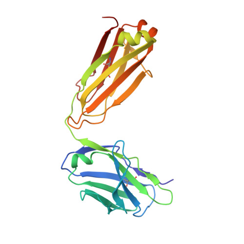
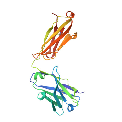
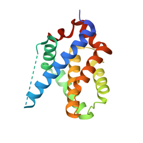
    BAX and BAK are pro-apoptotic members of the BCL2 family that are required to permeabilize the mitochondrial outer membrane. The proteins can adopt a non-activated monomeric conformation, or an activated conformation in which the exposed BH3 domain facilitates binding either to a prosurvival protein or to another activated BAK or BAX protein to promote pore formation. Certain cancer cells are proposed to have high levels of activated BAK sequestered by MCL1 or BCLX L , thus priming these cells to undergo apoptosis in response to BH3 mimetic compounds that target MCL1 or BCLX L . Here we report the first antibody, 14G6, that is specific for the non-activated BAK conformer. A crystal structure of 14G6 Fab bound to BAK revealed a binding site encompassing both the α1 helix and α5-α6 hinge regions of BAK, two sites involved in the unfolding of BAK during its activation. In mitochondrial experiments, 14G6 inhibited BAK unfolding triggered by three diverse BAK activators, supporting crucial roles for both α1 dissociation and separation of the core (α2-α5) and latch (α6-α9) regions in BAK activation. 14G6 bound the majority of BAK in several leukaemia cell lines, and binding decreased following treatment with BH3 mimetics, indicating only minor levels of constitutively activated BAK in those cells. In summary, 14G6 provides a new means of assessing BAK status in response to anti-cancer treatments.


  • Organizational Affiliation

    Walter and Eliza Hall Institute of Medical Research, 1G Royal Parade, Parkville, VIC, 3052, Australia.


Macromolecules
Find similar proteins by:  (by identity cutoff)  |  3D Structure
Entity ID: 1
MoleculeChains Sequence LengthOrganismDetailsImage
14G6 Fab light chainA [auth L],
D [auth A]
213Homo sapiensMutation(s): 0 
Entity Groups  
Sequence Clusters30% Identity50% Identity70% Identity90% Identity95% Identity100% Identity
Sequence Annotations
Expand
  • Reference Sequence
Find similar proteins by:  (by identity cutoff)  |  3D Structure
Entity ID: 2
MoleculeChains Sequence LengthOrganismDetailsImage
14G6 Fab heavy chainB [auth H],
E [auth B]
219Homo sapiensMutation(s): 0 
Entity Groups  
Sequence Clusters30% Identity50% Identity70% Identity90% Identity95% Identity100% Identity
Sequence Annotations
Expand
  • Reference Sequence
Find similar proteins by:  (by identity cutoff)  |  3D Structure
Entity ID: 3
MoleculeChains Sequence LengthOrganismDetailsImage
Bcl-2 homologous antagonist/killerC,
F [auth D]
170Homo sapiensMutation(s): 1 
Gene Names: BAK1BAKBCL2L7CDN1
UniProt & NIH Common Fund Data Resources
Find proteins for Q16611 (Homo sapiens)
Explore Q16611 
Go to UniProtKB:  Q16611
PHAROS:  Q16611
GTEx:  ENSG00000030110 
Entity Groups  
Sequence Clusters30% Identity50% Identity70% Identity90% Identity95% Identity100% Identity
UniProt GroupQ16611
Sequence Annotations
Expand
  • Reference Sequence
Small Molecules
Ligands 7 Unique
IDChains Name / Formula / InChI Key2D Diagram3D Interactions
1PE
Query on 1PE

Download Ideal Coordinates CCD File 
S [auth A]PENTAETHYLENE GLYCOL
C10 H22 O6
JLFNLZLINWHATN-UHFFFAOYSA-N
PGE
Query on PGE

Download Ideal Coordinates CCD File 
O [auth H],
P [auth H],
W [auth A]
TRIETHYLENE GLYCOL
C6 H14 O4
ZIBGPFATKBEMQZ-UHFFFAOYSA-N
144
Query on 144

Download Ideal Coordinates CCD File 
N [auth H],
Z [auth B]
TRIS-HYDROXYMETHYL-METHYL-AMMONIUM
C4 H12 N O3
DRDCQJADRSJFFD-UHFFFAOYSA-N
PEG
Query on PEG

Download Ideal Coordinates CCD File 
BA [auth D]
I [auth L]
L [auth H]
M [auth H]
Q [auth A]
BA [auth D],
I [auth L],
L [auth H],
M [auth H],
Q [auth A],
V [auth A],
X [auth B],
Y [auth B]
DI(HYDROXYETHYL)ETHER
C4 H10 O3
MTHSVFCYNBDYFN-UHFFFAOYSA-N
SO4
Query on SO4

Download Ideal Coordinates CCD File 
AA [auth B],
G [auth L],
U [auth A]
SULFATE ION
O4 S
QAOWNCQODCNURD-UHFFFAOYSA-L
EDO
Query on EDO

Download Ideal Coordinates CCD File 
R [auth A],
T [auth A]
1,2-ETHANEDIOL
C2 H6 O2
LYCAIKOWRPUZTN-UHFFFAOYSA-N
CCN
Query on CCN

Download Ideal Coordinates CCD File 
H [auth L],
J [auth L],
K [auth L]
ACETONITRILE
C2 H3 N
WEVYAHXRMPXWCK-UHFFFAOYSA-N
Experimental Data & Validation

Experimental Data

  • Method: X-RAY DIFFRACTION
  • Resolution: 2.40 Å
  • R-Value Free: 0.252 
  • R-Value Work: 0.215 
  • R-Value Observed: 0.217 
  • Space Group: P 1
Unit Cell:
Length ( Å )Angle ( ˚ )
a = 71.554α = 103.01
b = 72.622β = 103.98
c = 75.282γ = 112.15
Software Package:
Software NamePurpose
XDSdata reduction
XSCALEdata scaling
MOLREPphasing
PHENIXrefinement
PDB_EXTRACTdata extraction

Structure Validation

View Full Validation Report


Entry History & Funding Information

Deposition Data


Funding OrganizationLocationGrant Number
National Health and Medical Research Council (NHMRC, Australia)Australia1016701
Leukemia & Lymphoma SocietyUnited States--

Revision History  (Full details and data files)

  • Version 1.0: 2024-04-10
    Type: Initial release
  • Version 1.1: 2024-04-17
    Changes: Database references