8UFB

Eastern equine encephalitis virus (PE-6) VLP in complex with full-length VLDLR (asymmetric unit)


Experimental Data Snapshot

  • Method: ELECTRON MICROSCOPY
  • Resolution: 3.89 Å
  • Aggregation State: PARTICLE 
  • Reconstruction Method: SINGLE PARTICLE 

wwPDB Validation   3D Report Full Report


This is version 1.1 of the entry. See complete history


Literature

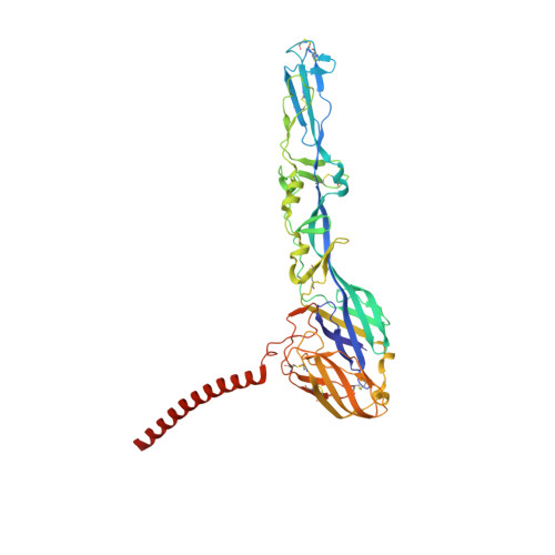
Structural and functional basis of VLDLR usage by Eastern equine encephalitis virus.

Adams, L.J.Raju, S.Ma, H.Gilliland Jr., T.Reed, D.S.Klimstra, W.B.Fremont, D.H.Diamond, M.S.

(2024) Cell 187: 360-374.e19

  • DOI: https://doi.org/10.1016/j.cell.2023.11.031
  • Primary Citation of Related Structures:  
    8UFA, 8UFB, 8UFC

  • PubMed Abstract: 

    The very-low-density lipoprotein receptor (VLDLR) comprises eight LDLR type A (LA) domains and supports entry of distantly related alphaviruses, including Eastern equine encephalitis virus (EEEV) and Semliki Forest virus (SFV). Here, by resolving multiple cryo-electron microscopy structures of EEEV-VLDLR complexes and performing mutagenesis and functional studies, we show that EEEV uses multiple sites (E1/E2 cleft and E2 A domain) to engage more than one LA domain simultaneously. However, no single LA domain is necessary or sufficient to support efficient EEEV infection. Whereas all EEEV strains show conservation of two VLDLR-binding sites, the EEEV PE-6 strain and a few other EEE complex members feature a single amino acid substitution that enables binding of LA domains to an additional site on the E2 B domain. These structural and functional analyses informed the design of a minimal VLDLR decoy receptor that neutralizes EEEV infection and protects mice from lethal challenge.


  • Organizational Affiliation

    Department of Pathology & Immunology, Washington University School of Medicine, St. Louis, MO 63110, USA.


Macromolecules
Find similar proteins by:  (by identity cutoff)  |  3D Structure
Entity ID: 1
MoleculeChains Sequence LengthOrganismDetailsImage
E1 protein
A, D, G, J
441Eastern equine encephalitis virusMutation(s): 0 
UniProt
Find proteins for Q88678 (Eastern equine encephalitis virus)
Explore Q88678 
Go to UniProtKB:  Q88678
Entity Groups  
Sequence Clusters30% Identity50% Identity70% Identity90% Identity95% Identity100% Identity
UniProt GroupQ88678
Sequence Annotations
Expand
  • Reference Sequence
Find similar proteins by:  (by identity cutoff)  |  3D Structure
Entity ID: 2
MoleculeChains Sequence LengthOrganismDetailsImage
E2 protein
B, E, H, K
414Eastern equine encephalitis virusMutation(s): 0 
UniProt
Find proteins for Q88678 (Eastern equine encephalitis virus)
Explore Q88678 
Go to UniProtKB:  Q88678
Entity Groups  
Sequence Clusters30% Identity50% Identity70% Identity90% Identity95% Identity100% Identity
UniProt GroupQ88678
Sequence Annotations
Expand
  • Reference Sequence
Find similar proteins by:  (by identity cutoff)  |  3D Structure
Entity ID: 3
MoleculeChains Sequence LengthOrganismDetailsImage
Capsid protein
C, F, I, L
151Eastern equine encephalitis virusMutation(s): 0 
Gene Names: SP
UniProt
Find proteins for W8S146 (Eastern equine encephalitis virus)
Explore W8S146 
Go to UniProtKB:  W8S146
Entity Groups  
Sequence Clusters30% Identity50% Identity70% Identity90% Identity95% Identity100% Identity
UniProt GroupW8S146
Sequence Annotations
Expand
  • Reference Sequence
Find similar proteins by:  (by identity cutoff)  |  3D Structure
Entity ID: 4
MoleculeChains Sequence LengthOrganismDetailsImage
Isoform Short of Very low-density lipoprotein receptor748Homo sapiensMutation(s): 0 
Gene Names: VLDLR
UniProt & NIH Common Fund Data Resources
Find proteins for P98155 (Homo sapiens)
Explore P98155 
Go to UniProtKB:  P98155
PHAROS:  P98155
GTEx:  ENSG00000147852 
Entity Groups  
Sequence Clusters30% Identity50% Identity70% Identity90% Identity95% Identity100% Identity
UniProt GroupP98155
Sequence Annotations
Expand
  • Reference Sequence
Small Molecules
Ligands 2 Unique
IDChains Name / Formula / InChI Key2D Diagram3D Interactions
NAG
Query on NAG

Download Ideal Coordinates CCD File 
AA [auth J]
BA [auth K]
U [auth A]
V [auth B]
W [auth D]
AA [auth J],
BA [auth K],
U [auth A],
V [auth B],
W [auth D],
X [auth E],
Y [auth G],
Z [auth H]
2-acetamido-2-deoxy-beta-D-glucopyranose
C8 H15 N O6
OVRNDRQMDRJTHS-FMDGEEDCSA-N
CA
Query on CA

Download Ideal Coordinates CCD File 
CA [auth V]
DA [auth V]
EA [auth W]
FA [auth W]
GA [auth X]
CA [auth V],
DA [auth V],
EA [auth W],
FA [auth W],
GA [auth X],
HA [auth X],
IA [auth Y],
JA [auth Y],
KA [auth R],
LA [auth S],
MA [auth T],
NA [auth U]
CALCIUM ION
Ca
BHPQYMZQTOCNFJ-UHFFFAOYSA-N
Experimental Data & Validation

Experimental Data

  • Method: ELECTRON MICROSCOPY
  • Resolution: 3.89 Å
  • Aggregation State: PARTICLE 
  • Reconstruction Method: SINGLE PARTICLE 

Structure Validation

View Full Validation Report


Entry History & Funding Information

Deposition Data


Funding OrganizationLocationGrant Number
National Institutes of Health/National Institute Of Allergy and Infectious Diseases (NIH/NIAID)United States--

Revision History  (Full details and data files)

  • Version 1.0: 2024-01-17
    Type: Initial release
  • Version 1.1: 2024-01-31
    Changes: Database references