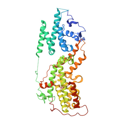
8UF2

Apo SOS2 crystal structure in P1 space group


Experimental Data Snapshot

  • Method: X-RAY DIFFRACTION
  • Resolution: 1.60 Å
  • R-Value Free: 0.232 
  • R-Value Work: 0.196 
  • R-Value Observed: 0.198 

wwPDB Validation   3D Report Full Report


This is version 1.1 of the entry. See complete history


Literature

Discovery of Five SOS2 Fragment Hits with Binding Modes Determined by SOS2 X-Ray Cocrystallography.

Smith, C.R.Chen, D.Christensen, J.G.Coulombe, R.Fethiere, J.Gunn, R.J.Hollander, J.Jones, B.Ketcham, J.M.Khare, S.Kuehler, J.Lawson, J.D.Marx, M.A.Olson, P.Pearson, K.E.Ren, C.Tsagris, D.Ulaganathan, T.Van't Veer, I.Wang, X.Ivetac, A.

(2024) J Med Chem 67: 774-781

  • DOI: https://doi.org/10.1021/acs.jmedchem.3c02140
  • Primary Citation of Related Structures:  
    8T5G, 8T5M, 8T5R, 8UC9, 8UF2, 8UH0

  • PubMed Abstract: 

    SOS1 and SOS2 are guanine nucleotide exchange factors that mediate RTK-stimulated RAS activation. Selective SOS1:KRAS PPI inhibitors are currently under clinical investigation, whereas there are no reports to date of SOS2:KRAS PPI inhibitors. SOS2 activity is implicated in MAPK rebound when divergent SOS1 mutant cell lines are treated with the SOS1 inhibitor BI-3406; therefore, SOS2:KRAS inhibitors are of therapeutic interest. In this report, we detail a fragment-based screening strategy to identify X-ray cocrystal structures of five diverse fragment hits bound to SOS2.


  • Organizational Affiliation

    Mirati Therapeutics, San Diego, California 92130, United States.


Macromolecules
Find similar proteins by:  (by identity cutoff)  |  3D Structure
Entity ID: 1
MoleculeChains Sequence LengthOrganismDetailsImage
Son of sevenless homolog 2490Homo sapiensMutation(s): 0 
Gene Names: SOS2
UniProt & NIH Common Fund Data Resources
Find proteins for Q07890 (Homo sapiens)
Explore Q07890 
Go to UniProtKB:  Q07890
PHAROS:  Q07890
GTEx:  ENSG00000100485 
Entity Groups  
Sequence Clusters30% Identity50% Identity70% Identity90% Identity95% Identity100% Identity
UniProt GroupQ07890
Sequence Annotations
Expand
  • Reference Sequence
Small Molecules
Ligands 1 Unique
IDChains Name / Formula / InChI Key2D Diagram3D Interactions
SO4
Query on SO4

Download Ideal Coordinates CCD File 
B [auth A]SULFATE ION
O4 S
QAOWNCQODCNURD-UHFFFAOYSA-L
Experimental Data & Validation

Experimental Data

  • Method: X-RAY DIFFRACTION
  • Resolution: 1.60 Å
  • R-Value Free: 0.232 
  • R-Value Work: 0.196 
  • R-Value Observed: 0.198 
  • Space Group: P 1
Unit Cell:
Length ( Å )Angle ( ˚ )
a = 46.665α = 85.581
b = 52.002β = 75.304
c = 61.865γ = 76.949
Software Package:
Software NamePurpose
PHENIXrefinement
HKL-2000data reduction
HKL-2000data scaling
PHASERphasing

Structure Validation

View Full Validation Report


Entry History & Funding Information

Deposition Data


Funding OrganizationLocationGrant Number
Not fundedUnited States--

Revision History  (Full details and data files)

  • Version 1.0: 2024-01-10
    Type: Initial release
  • Version 1.1: 2024-01-24
    Changes: Database references