8UAP

Crystal Structure of Human G Protein-Coupled Receptor Kinase 5 D311N in Complex with CCG273441


Experimental Data Snapshot

  • Method: X-RAY DIFFRACTION
  • Resolution: 2.50 Å
  • R-Value Free: 0.267 
  • R-Value Work: 0.227 
  • R-Value Observed: 0.229 

wwPDB Validation   3D Report Full Report


Ligand Structure Quality Assessment 


This is version 1.0 of the entry. See complete history


Literature

Development of a new class of potent and highly selective G protein-coupled receptor kinase 5 inhibitors and structural insight from crystal structures of inhibitor complexes.

Chen, Y.Sonawane, A.Manda, R.Gadi, R.K.Tesmer, J.J.G.Ghosh, A.K.

(2023) Eur J Med Chem 264: 115931-115931

  • DOI: https://doi.org/10.1016/j.ejmech.2023.115931
  • Primary Citation of Related Structures:  
    8UAP, 8UAQ

  • PubMed Abstract: 

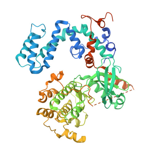
    G protein-coupled receptor kinase 5 (GRK5) is an important drug development target for heart failure, cardiac hypertrophy, and cancer. We have designed and developed a new class of highly selective, potent, and non-covalent GRK5 inhibitors. One of the inhibitors displayed GRK5 IC 50 value of 10 nM and exhibited >100,000-fold selectivity over GRK2. The X-ray structure of a ketoamide-derived inhibitor-bound GRK5 showed the formation of a hemithioketal intermediate with active site Cys474 in the GRK5 active site and provided new insights into the ligand-binding site interactions responsible for high selectivity. The current studies serve as an important guide to therapeutic GRK5 inhibitor drug development.


  • Organizational Affiliation

    Department of Biological Sciences, Purdue University, West Lafayette, IN, 47907, USA; Department of Medicinal Chemistry and Molecular Pharmacology, Purdue University, West Lafayette, IN, 47907, USA.


Macromolecules
Find similar proteins by:  (by identity cutoff)  |  3D Structure
Entity ID: 1
MoleculeChains Sequence LengthOrganismDetailsImage
G protein-coupled receptor kinase 5598Homo sapiensMutation(s): 1 
Gene Names: GRK5
UniProt & NIH Common Fund Data Resources
Find proteins for P34947 (Homo sapiens)
Explore P34947 
Go to UniProtKB:  P34947
PHAROS:  P34947
GTEx:  ENSG00000198873 
Entity Groups  
Sequence Clusters30% Identity50% Identity70% Identity90% Identity95% Identity100% Identity
UniProt GroupP34947
Sequence Annotations
Expand
  • Reference Sequence
Small Molecules
Ligands 1 Unique
IDChains Name / Formula / InChI Key2D Diagram3D Interactions
W2T
Query on W2T

Download Ideal Coordinates CCD File 
B [auth A](3Z)-3-{[4-(2-chloroacetamido)-3,5-dimethyl-1H-pyrrol-2-yl]methylidene}-N-[(1R)-1-(4-fluorophenyl)ethyl]-2-oxo-2,3-dihydro-1H-indole-5-carboxamide
C26 H24 Cl F N4 O3
IDWBLFLYQVHAFH-LPKQHDGYSA-N
Experimental Data & Validation

Experimental Data

  • Method: X-RAY DIFFRACTION
  • Resolution: 2.50 Å
  • R-Value Free: 0.267 
  • R-Value Work: 0.227 
  • R-Value Observed: 0.229 
  • Space Group: P 41 21 2
Unit Cell:
Length ( Å )Angle ( ˚ )
a = 137.855α = 90
b = 137.855β = 90
c = 71.089γ = 90
Software Package:
Software NamePurpose
PHENIXrefinement
xia2data reduction
xia2data scaling
MOLREPphasing

Structure Validation

View Full Validation Report


Ligand Structure Quality Assessment 


Entry History & Funding Information

Deposition Data


Funding OrganizationLocationGrant Number
National Institutes of Health/National Heart, Lung, and Blood Institute (NIH/NHLBI)United StatesHL071818
National Institutes of Health/National Center for Research Resources (NIH/NCRR)United StatesCA023168

Revision History  (Full details and data files)

  • Version 1.0: 2023-12-13
    Type: Initial release