8U8U | pdb_00008u8u

Cryo-EM Structure of Cognate Substrate ATP Bound in the Entry Site (ES) of Human Mitochondrial Transcription Elongation Complex


Experimental Data Snapshot

  • Method: ELECTRON MICROSCOPY
  • Resolution: 2.90 Å
  • Aggregation State: PARTICLE 
  • Reconstruction Method: SINGLE PARTICLE 

Starting Model: experimental
View more details

wwPDB Validation 3D Report Full Report

Validation slider image for 8U8U

This is version 1.0 of the entry. See complete history

Literature

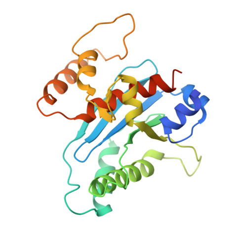
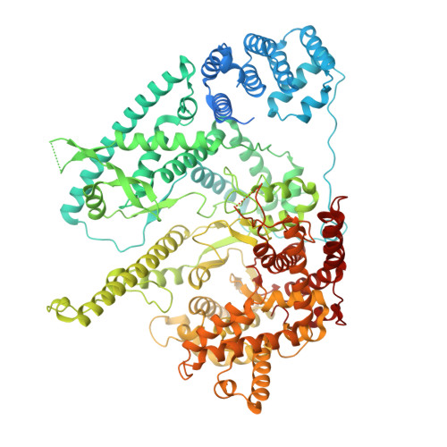
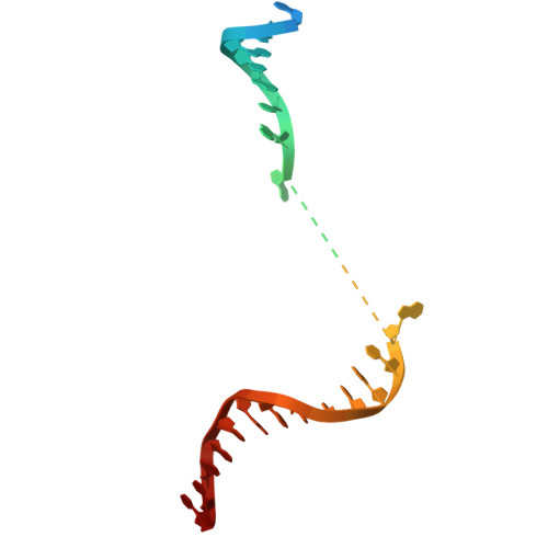
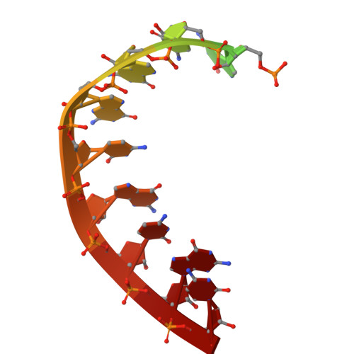
Structural basis for substrate binding and selection by human mitochondrial RNA polymerase.

Herbine, K.Nayak, A.R.Temiakov, D.

(2024) Nat Commun 15: 7134-7134

  • DOI: https://doi.org/10.1038/s41467-024-50817-9
  • Primary Citation Related Structures: 
    8U8U, 8U8V, 9BDC, 9BDD

  • PubMed Abstract: 

    The mechanism by which RNAP selects cognate substrates and discriminates between deoxy and ribonucleotides is of fundamental importance to the fidelity of transcription. Here, we present cryo-EM structures of human mitochondrial transcription elongation complexes that reveal substrate ATP bound in Entry and Insertion Sites. In the Entry Site, the substrate binds along the O helix of the fingers domain of mtRNAP but does not interact with the templating DNA base. Interactions between RNAP and the triphosphate moiety of the NTP in the Entry Site ensure discrimination against nucleosides and their diphosphate and monophosphate derivatives but not against non-cognate rNTPs and dNTPs. Closing of the fingers domain over the catalytic site results in delivery of both the templating DNA base and the substrate into the Insertion Site and recruitment of the catalytic magnesium ions. The cryo-EM data also reveal a conformation adopted by mtRNAP to reject a non-cognate substrate from its active site. Our findings establish a structural basis for substrate binding and suggest a unified mechanism of NTP selection for single-subunit RNAPs.


  • Organizational Affiliation
    • Department of Biochemistry and Molecular Biology, Thomas Jefferson University, 1020 Locust St, Philadelphia, PA, 19107, USA.

Macromolecule Content 

  • Total Structure Weight: 207.58 kDa 
  • Atom Count: 12,344 
  • Modeled Residue Count: 1,459 
  • Deposited Residue Count: 1,665 
  • Unique protein chains: 2
  • Unique nucleic acid chains: 3

Macromolecules


Find similar proteins by:|  3D Structure
Entity ID: 1
MoleculeChains  Sequence LengthOrganismDetailsImage
Transcription elongation factor, mitochondrial
A, B
232Homo sapiensMutation(s): 0 
Gene Names: TEFMC17orf42
UniProt & NIH Common Fund Data Resources
Find proteins for Q96QE5 (Homo sapiens)
Explore Q96QE5 
Go to UniProtKB:  Q96QE5
PHAROS:  Q96QE5
GTEx:  ENSG00000172171 
Entity Groups
Sequence Clusters30% Identity50% Identity70% Identity90% Identity95% Identity100% Identity
UniProt GroupQ96QE5
Sequence Annotations
Expand
Reference Sequence
Find similar proteins by:|  3D Structure
Entity ID: 2
MoleculeChains  Sequence LengthOrganismDetailsImage
DNA-directed RNA polymerase, mitochondrialC [auth E]1,119Homo sapiensMutation(s): 0 
Gene Names: POLRMT
EC: 2.7.7.6
UniProt & NIH Common Fund Data Resources
Find proteins for O00411 (Homo sapiens)
Explore O00411 
Go to UniProtKB:  O00411
PHAROS:  O00411
GTEx:  ENSG00000099821 
Entity Groups
Sequence Clusters30% Identity50% Identity70% Identity90% Identity95% Identity100% Identity
UniProt GroupO00411
Sequence Annotations
Expand
Reference Sequence
Find similar nucleic acids by:  (by identity cutoff) 
Entity ID: 3
MoleculeChains LengthOrganismImage
Non-Template Strand DNA (NT27mt)D [auth N]34synthetic construct
Sequence Annotations
Expand
Reference Sequence
Find similar nucleic acids by:  Sequence
Entity ID: 4
MoleculeChains LengthOrganismImage
RNA (RNA14mt)E [auth R]14synthetic construct
Sequence Annotations
Expand
Reference Sequence
Find similar nucleic acids by:  (by identity cutoff) 
Entity ID: 5
MoleculeChains LengthOrganismImage
Template Strand DNA (TS31mt)F [auth T]34synthetic construct
Sequence Annotations
Expand
Reference Sequence

Small Molecules

Ligands 1 Unique
IDChains Name / Formula / InChI Key2D Diagram3D Interactions
APC

Query on APC


Download:Ideal Coordinates CCD File
G [auth E]DIPHOSPHOMETHYLPHOSPHONIC ACID ADENOSYL ESTER
C11 H18 N5 O12 P3
CAWZRIXWFRFUQB-IOSLPCCCSA-N

Experimental Data & Validation

Experimental Data

  • Method: ELECTRON MICROSCOPY
  • Resolution: 2.90 Å
  • Aggregation State: PARTICLE 
  • Reconstruction Method: SINGLE PARTICLE 
EM Software:
TaskSoftware PackageVersion
MODEL REFINEMENTPHENIX1.21rc1_5015:

Structure Validation

View Full Validation Report


Entry History 

& Funding Information

Deposition Data


Funding OrganizationLocationGrant Number
National Institutes of Health/National Institute of General Medical Sciences (NIH/NIGMS)United StatesNIH GM131832

Revision History  (Full details and data files)

  • Version 1.0: 2024-09-04
    Type: Initial release