8U5J | pdb_00008u5j

Structure of Mango III variant aptamer bound to T01-07M-B


Experimental Data Snapshot

  • Method: X-RAY DIFFRACTION
  • Resolution: 1.70 Å
  • R-Value Free: 
    0.236 (Depositor), 0.238 (DCC) 
  • R-Value Work: 
    0.201 (Depositor), 0.202 (DCC) 
  • R-Value Observed: 
    0.205 (Depositor) 

Starting Model: experimental
View more details

wwPDB Validation 3D Report Full Report

Validation slider image for 8U5J

Ligand Structure Quality Assessment 


This is version 1.1 of the entry. See complete history

Literature

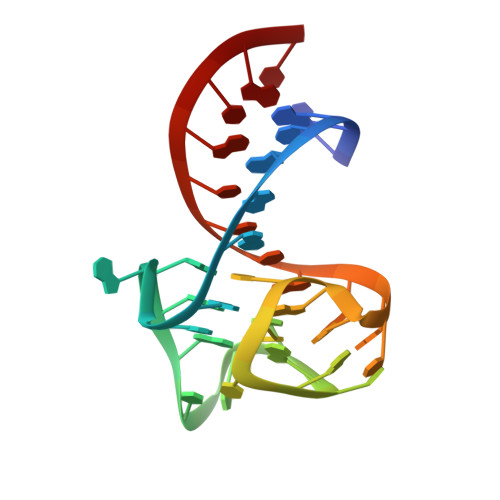
Symmetry breaking of fluorophore binding to a G-quadruplex generates an RNA aptamer with picomolar KD.

Lu, X.Passalacqua, L.F.M.Nodwell, M.Kong, K.Y.S.Caballero-Garcia, G.Dolgosheina, E.V.Ferre-D'Amare, A.R.Britton, R.Unrau, P.J.

(2024) Nucleic Acids Res 52: 8039-8051

  • DOI: https://doi.org/10.1093/nar/gkae493
  • Primary Citation Related Structures: 
    8U5J, 8U5K, 8U5P, 8U5R, 8U5T, 8U5Z, 8U60

  • PubMed Abstract: 

    Fluorogenic RNA aptamer tags with high affinity enable RNA purification and imaging. The G-quadruplex (G4) based Mango (M) series of aptamers were selected to bind a thiazole orange based (TO1-Biotin) ligand. Using a chemical biology and reselection approach, we have produced a MII.2 aptamer-ligand complex with a remarkable set of properties: Its unprecedented KD of 45 pM, formaldehyde resistance (8% v/v), temperature stability and ligand photo-recycling properties are all unusual to find simultaneously within a small RNA tag. Crystal structures demonstrate how MII.2, which differs from MII by a single A23U mutation, and modification of the TO1-Biotin ligand to TO1-6A-Biotin achieves these results. MII binds TO1-Biotin heterogeneously via a G4 surface that is surrounded by a stadium of five adenosines. Breaking this pseudo-rotational symmetry results in a highly cooperative and homogeneous ligand binding pocket: A22 of the G4 stadium stacks on the G4 binding surface while the TO1-6A-Biotin ligand completely fills the remaining three quadrants of the G4 ligand binding face. Similar optimization attempts with MIII.1, which already binds TO1-Biotin in a homogeneous manner, did not produce such marked improvements. We use the novel features of the MII.2 complex to demonstrate a powerful optically-based RNA purification system.


  • Organizational Affiliation
    • Department of Molecular Biology and Biochemistry, Simon Fraser University, Burnaby, British Columbia V5A 1S6, Canada.

Macromolecule Content 

  • Total Structure Weight: 12.65 kDa 
  • Atom Count: 987 
  • Modeled Residue Count: 36 
  • Deposited Residue Count: 36 
  • Unique nucleic acid chains: 1

Macromolecules

Find similar nucleic acids by:  (by identity cutoff) 
Entity ID: 1
MoleculeChains LengthOrganismImage
Mango III variant36synthetic construct
Sequence Annotations
Expand
Reference Sequence

Small Molecules

Ligands 3 Unique
IDChains Name / Formula / InChI Key2D Diagram3D Interactions
W6F
(Subject of Investigation/LOI)

Query on W6F


Download:Ideal Coordinates CCD File
E [auth A]2-[(E)-(1,7-dimethylquinolin-4(1H)-ylidene)methyl]-3-{2,16-dioxo-20-[(3aR,4S,6aR)-2-oxohexahydro-1H-thieno[3,4-d]imidazol-4-yl]-6,9,12-trioxa-3,15-diazaicosan-1-yl}-1,3-benzothiazol-3-ium
C39 H51 N6 O6 S2
OPDJCBUXWAHTAM-PJXAABHXSA-O
DMS

Query on DMS


Download:Ideal Coordinates CCD File
C [auth A],
D [auth A]
DIMETHYL SULFOXIDE
C2 H6 O S
IAZDPXIOMUYVGZ-UHFFFAOYSA-N
K

Query on K


Download:Ideal Coordinates CCD File
B [auth A]POTASSIUM ION
K
NPYPAHLBTDXSSS-UHFFFAOYSA-N

Experimental Data & Validation

Experimental Data

  • Method: X-RAY DIFFRACTION
  • Resolution: 1.70 Å
  • R-Value Free:  0.236 (Depositor), 0.238 (DCC) 
  • R-Value Work:  0.201 (Depositor), 0.202 (DCC) 
  • R-Value Observed: 0.205 (Depositor) 
Space Group: P 32 2 1
Unit Cell:
Length ( Å )Angle ( ˚ )
a = 55.66α = 90
b = 55.66β = 90
c = 65.715γ = 120
Software Package:
Software NamePurpose
PHENIXrefinement
PHASERphasing
DIALSdata reduction
DIALSdata scaling

Structure Validation

View Full Validation Report


Ligand Structure Quality Assessment 


Entry History 

& Funding Information

Deposition Data


Funding OrganizationLocationGrant Number
National Institutes of Health/National Heart, Lung, and Blood Institute (NIH/NHLBI)United States--

Revision History  (Full details and data files)

  • Version 1.0: 2024-03-27
    Type: Initial release
  • Version 1.1: 2025-10-08
    Changes: Database references, Structure summary