8U3D | pdb_00008u3d

Structure of Apo Sialin at pH7.5

  • Classification: MEMBRANE PROTEIN
  • Organism(s): Homo sapiens
  • Expression System: Homo sapiens
  • Mutation(s): No 
  • Membrane Protein: Yes  PDBTMMemProtMDmpstruc

  • Deposited: 2023-09-07 Released: 2024-05-29 
  • Deposition Author(s): Schmiege, P., Li, X.
  • Funding Organization(s): National Institutes of Health/National Institute of General Medical Sciences (NIH/NIGMS), National Institutes of Health/Eunice Kennedy Shriver National Institute of Child Health & Human Development (NIH/NICHD), Welch Foundation

Experimental Data Snapshot

  • Method: ELECTRON MICROSCOPY
  • Resolution: 2.83 Å
  • Aggregation State: PARTICLE 
  • Reconstruction Method: SINGLE PARTICLE 

wwPDB Validation 3D Report Full Report

Validation slider image for 8U3D

This is version 1.1 of the entry. See complete history

Literature

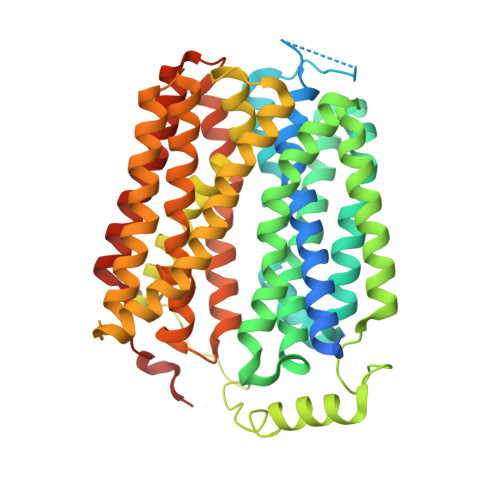
Structure and inhibition of the human lysosomal transporter Sialin.

Schmiege, P.Donnelly, L.Elghobashi-Meinhardt, N.Lee, C.H.Li, X.

(2024) Nat Commun 15: 4386-4386

  • DOI: https://doi.org/10.1038/s41467-024-48535-3
  • Primary Citation Related Structures: 
    8U3D, 8U3E, 8U3F, 8U3G, 8U3H, 9AYB

  • PubMed Abstract: 

    Sialin, a member of the solute carrier 17 (SLC17) transporter family, is unique in its ability to transport not only sialic acid using a pH-driven mechanism, but also transport mono and diacidic neurotransmitters, such as glutamate and N-acetylaspartylglutamate (NAAG), into synaptic vesicles via a membrane potential-driven mechanism. While most transporters utilize one of these mechanisms, the structural basis of how Sialin transports substrates using both remains unclear. Here, we present the cryogenic electron-microscopy structures of human Sialin: apo cytosol-open, apo lumen-open, NAAG-bound, and inhibitor-bound. Our structures show that a positively charged cytosol-open vestibule accommodates either NAAG or the Sialin inhibitor Fmoc-Leu-OH, while its luminal cavity potentially binds sialic acid. Moreover, functional analyses along with molecular dynamics simulations identify key residues in binding sialic acid and NAAG. Thus, our findings uncover the essential conformational states in NAAG and sialic acid transport, demonstrating a working model of SLC17 transporters.


  • Organizational Affiliation
    • Department of Molecular Genetics, University of Texas Southwestern Medical Center, Dallas, TX, USA.

Macromolecule Content 

  • Total Structure Weight: 55.68 kDa 
  • Atom Count: 3,338 
  • Modeled Residue Count: 427 
  • Deposited Residue Count: 503 
  • Unique protein chains: 1

Macromolecules

Find similar proteins by:|  3D Structure
Entity ID: 1
MoleculeChains  Sequence LengthOrganismDetailsImage
Sialin503Homo sapiensMutation(s): 0 
Gene Names: SLC17A5
Membrane Entity: Yes 
UniProt & NIH Common Fund Data Resources
Find proteins for Q9NRA2 (Homo sapiens)
Explore Q9NRA2 
Go to UniProtKB:  Q9NRA2
PHAROS:  Q9NRA2
GTEx:  ENSG00000119899 
Entity Groups
Sequence Clusters30% Identity50% Identity70% Identity90% Identity95% Identity100% Identity
UniProt GroupQ9NRA2
Sequence Annotations
Expand
Reference Sequence

Experimental Data & Validation

Experimental Data

  • Method: ELECTRON MICROSCOPY
  • Resolution: 2.83 Å
  • Aggregation State: PARTICLE 
  • Reconstruction Method: SINGLE PARTICLE 
EM Software:
TaskSoftware PackageVersion
MODEL REFINEMENTPHENIX1.20.1-4487
MODEL REFINEMENTISOLDE1.6.0

Structure Validation

View Full Validation Report


Entry History 

& Funding Information

Deposition Data


Funding OrganizationLocationGrant Number
National Institutes of Health/National Institute of General Medical Sciences (NIH/NIGMS)United States135343
National Institutes of Health/Eunice Kennedy Shriver National Institute of Child Health & Human Development (NIH/NICHD)United States110229
Welch FoundationUnited StatesI-1957

Revision History  (Full details and data files)

  • Version 1.0: 2024-05-29
    Type: Initial release
  • Version 1.1: 2024-12-11
    Changes: Data collection, Database references, Structure summary