8U1L

Cryo-EM structure of the RAF1-HSP90-CDC37 complex in the closed state


Experimental Data Snapshot

  • Method: ELECTRON MICROSCOPY
  • Resolution: 3.70 Å
  • Aggregation State: PARTICLE 
  • Reconstruction Method: SINGLE PARTICLE 

wwPDB Validation   3D Report Full Report


This is version 1.0 of the entry. See complete history


Literature

Structural dynamics of RAF1-HSP90-CDC37 and HSP90 complexes reveal asymmetric client interactions and key structural elements.

Finci, L.I.Chakrabarti, M.Gulten, G.Finney, J.Grose, C.Fox, T.Yang, R.Nissley, D.V.McCormick, F.Esposito, D.Balius, T.E.Simanshu, D.K.

(2024) Commun Biol 7: 260-260

  • DOI: https://doi.org/10.1038/s42003-024-05959-3
  • Primary Citation of Related Structures:  
    8U1L, 8U1M, 8U1N

  • PubMed Abstract: 

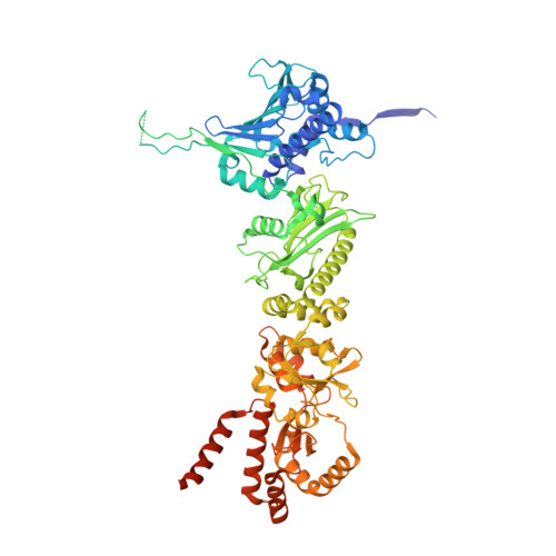
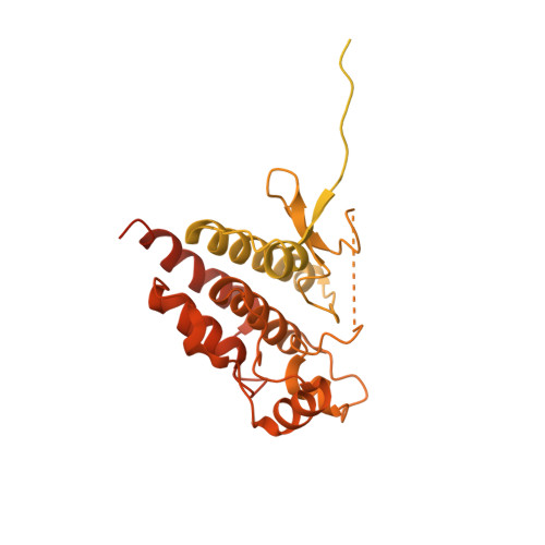
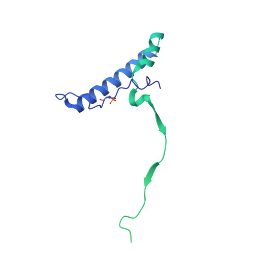
    RAF kinases are integral to the RAS-MAPK signaling pathway, and proper RAF1 folding relies on its interaction with the chaperone HSP90 and the cochaperone CDC37. Understanding the intricate molecular interactions governing RAF1 folding is crucial for comprehending this process. Here, we present a cryo-EM structure of the closed-state RAF1-HSP90-CDC37 complex, where the C-lobe of the RAF1 kinase domain binds to one side of the HSP90 dimer, and an unfolded N-lobe segment of the RAF1 kinase domain threads through the center of the HSP90 dimer. CDC37 binds to the kinase C-lobe, mimicking the N-lobe with its HxNI motif. We also describe structures of HSP90 dimers without RAF1 and CDC37, displaying only N-terminal and middle domains, which we term the semi-open state. Employing 1 μs atomistic simulations, energetic decomposition, and comparative structural analysis, we elucidate the dynamics and interactions within these complexes. Our quantitative analysis reveals that CDC37 bridges the HSP90-RAF1 interaction, RAF1 binds HSP90 asymmetrically, and that HSP90 structural elements engage RAF1's unfolded region. Additionally, N- and C-terminal interactions stabilize HSP90 dimers, and molecular interactions in HSP90 dimers rearrange between the closed and semi-open states. Our findings provide valuable insight into the contributions of HSP90 and CDC37 in mediating client folding.


  • Organizational Affiliation

    NCI RAS Initiative, Cancer Research Technology Program, Frederick National Laboratory for Cancer Research, Frederick, MD, USA.


Macromolecules
Find similar proteins by:  (by identity cutoff)  |  3D Structure
Entity ID: 1
MoleculeChains Sequence LengthOrganismDetailsImage
Heat shock protein 83
A, B
722Trichoplusia niMutation(s): 0 
UniProt
Find proteins for A0A7E5VSK5 (Trichoplusia ni)
Explore A0A7E5VSK5 
Go to UniProtKB:  A0A7E5VSK5
Entity Groups  
Sequence Clusters30% Identity50% Identity70% Identity90% Identity95% Identity100% Identity
UniProt GroupA0A7E5VSK5
Sequence Annotations
Expand
  • Reference Sequence
Find similar proteins by:  (by identity cutoff)  |  3D Structure
Entity ID: 2
MoleculeChains Sequence LengthOrganismDetailsImage
RAF proto-oncogene serine/threonine-protein kinase656Homo sapiensMutation(s): 0 
Gene Names: RAF1
UniProt & NIH Common Fund Data Resources
Find proteins for P04049 (Homo sapiens)
Explore P04049 
Go to UniProtKB:  P04049
PHAROS:  P04049
GTEx:  ENSG00000132155 
Entity Groups  
Sequence Clusters30% Identity50% Identity70% Identity90% Identity95% Identity100% Identity
UniProt GroupP04049
Sequence Annotations
Expand
  • Reference Sequence
Find similar proteins by:  (by identity cutoff)  |  3D Structure
Entity ID: 3
MoleculeChains Sequence LengthOrganismDetailsImage
Hsp90 co-chaperone Cdc37, N-terminally processed378Homo sapiensMutation(s): 0 
Gene Names: CDC37CDC37A
UniProt & NIH Common Fund Data Resources
Find proteins for Q16543 (Homo sapiens)
Explore Q16543 
Go to UniProtKB:  Q16543
PHAROS:  Q16543
GTEx:  ENSG00000105401 
Entity Groups  
Sequence Clusters30% Identity50% Identity70% Identity90% Identity95% Identity100% Identity
UniProt GroupQ16543
Sequence Annotations
Expand
  • Reference Sequence
Experimental Data & Validation

Experimental Data

  • Method: ELECTRON MICROSCOPY
  • Resolution: 3.70 Å
  • Aggregation State: PARTICLE 
  • Reconstruction Method: SINGLE PARTICLE 
EM Software:
TaskSoftware PackageVersion
RECONSTRUCTIONRELION3.1.3

Structure Validation

View Full Validation Report


Entry History & Funding Information

Deposition Data


Funding OrganizationLocationGrant Number
National Institutes of Health/National Cancer Institute (NIH/NCI)United StatesHHSN261200800001E

Revision History  (Full details and data files)

  • Version 1.0: 2024-03-13
    Type: Initial release