8TVC | pdb_00008tvc

Crystal structure of rA3G-ssDNA-AA


Experimental Data Snapshot

  • Method: X-RAY DIFFRACTION
  • Resolution: 1.93 Å
  • R-Value Free: 
    0.208 (Depositor), 0.211 (DCC) 
  • R-Value Work: 
    0.178 (Depositor), 0.178 (DCC) 
  • R-Value Observed: 
    0.179 (Depositor) 

Starting Model: experimental
View more details

wwPDB Validation 3D Report Full Report

Validation slider image for 8TVC

This is version 1.0 of the entry. See complete history

Literature

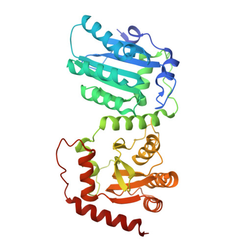
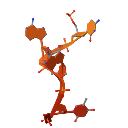
Molecular mechanism for regulating APOBEC3G DNA editing function by the non-catalytic domain.

Yang, H.Pacheco, J.Kim, K.Bokani, A.Ito, F.Ebrahimi, D.Chen, X.S.

(2024) Nat Commun 15: 8773-8773

  • DOI: https://doi.org/10.1038/s41467-024-52671-1
  • Primary Citation Related Structures: 
    8TVC, 8TX4

  • PubMed Abstract: 

    APOBEC3G, part of the AID/APOBEC cytidine deaminase family, is crucial for antiviral immunity. It has two zinc-coordinated cytidine-deaminase domains. The non-catalytic N-terminal domain strongly binds to nucleic acids, whereas the C-terminal domain catalyzes C-to-U editing in single-stranded DNA. The interplay between the two domains is not fully understood. Here, we show that DNA editing function of rhesus macaque APOBEC3G on linear and hairpin loop DNA is enhanced by AA or GA dinucleotide motifs present downstream in the 3'-direction of the target-C editing sites. The effective distance between AA/GA and the target-C sites is contingent on the local DNA secondary structure. We present two co-crystal structures of rhesus macaque APOBEC3G bound to ssDNA containing AA and GA, revealing the contribution of the non-catalytic domain in capturing AA/GA DNA. Our findings elucidate the molecular mechanism of APOBEC3G's cooperative function, which is critical for its antiviral role and its contribution to mutations in cancer genomes.


  • Organizational Affiliation
    • Molecular and Computational Biology, Departments of Biological Sciences, University of Southern California, Los Angeles, CA, 90089, USA.

Macromolecule Content 

  • Total Structure Weight: 51.8 kDa 
  • Atom Count: 3,470 
  • Modeled Residue Count: 379 
  • Deposited Residue Count: 407 
  • Unique protein chains: 1
  • Unique nucleic acid chains: 1

Macromolecules


Find similar proteins by:|  3D Structure
Entity ID: 1
MoleculeChains  Sequence LengthOrganismDetailsImage
DNA dC->dU-editing enzyme APOBEC-3G386Macaca mulattaMutation(s): 0 
Gene Names: APOBEC3G
EC: 3.5.4 (PDB Primary Data), 3.5.4.38 (UniProt)
UniProt
Find proteins for Q7YR23 (Macaca mulatta)
Explore Q7YR23 
Go to UniProtKB:  Q7YR23
Entity Groups
Sequence Clusters30% Identity50% Identity70% Identity90% Identity95% Identity100% Identity
UniProt GroupQ7YR23
Sequence Annotations
Expand
Reference Sequence
Find similar nucleic acids by:  Sequence
Entity ID: 2
MoleculeChains LengthOrganismImage
DNA 21-mer21synthetic construct
Sequence Annotations
Expand
Reference Sequence

Experimental Data & Validation

Experimental Data

  • Method: X-RAY DIFFRACTION
  • Resolution: 1.93 Å
  • R-Value Free:  0.208 (Depositor), 0.211 (DCC) 
  • R-Value Work:  0.178 (Depositor), 0.178 (DCC) 
  • R-Value Observed: 0.179 (Depositor) 
Space Group: P 21 21 21
Unit Cell:
Length ( Å )Angle ( ˚ )
a = 55.429α = 90
b = 68.461β = 90
c = 126.735γ = 90
Software Package:
Software NamePurpose
PHENIXrefinement
JBluIce-EPICSdata collection
PHASERphasing
xia2data scaling
xia2data reduction
Cootmodel building

Structure Validation

View Full Validation Report


Entry History 

& Funding Information

Deposition Data


Funding OrganizationLocationGrant Number
National Institutes of Health/National Institute Of Allergy and Infectious Diseases (NIH/NIAID)United States--

Revision History  (Full details and data files)

  • Version 1.0: 2024-11-06
    Type: Initial release