8TUI | pdb_00008tui

Crystal structure of Fab-Lirilumab bound to KIR2DL3


Experimental Data Snapshot

  • Method: X-RAY DIFFRACTION
  • Resolution: 2.75 Å
  • R-Value Free: 
    0.254 (Depositor), 0.253 (DCC) 
  • R-Value Work: 
    0.211 (Depositor), 0.210 (DCC) 
  • R-Value Observed: 
    0.213 (Depositor) 

Starting Models: experimental
View more details

wwPDB Validation 3D Report Full Report

Validation slider image for 8TUI

This is version 1.2 of the entry. See complete history

Literature

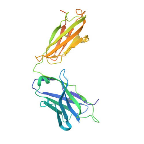
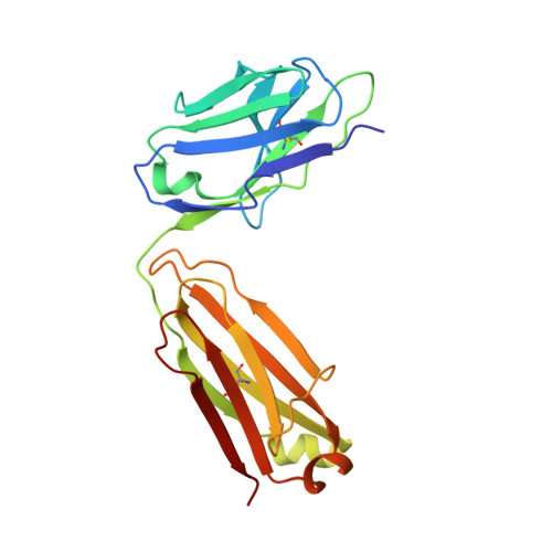
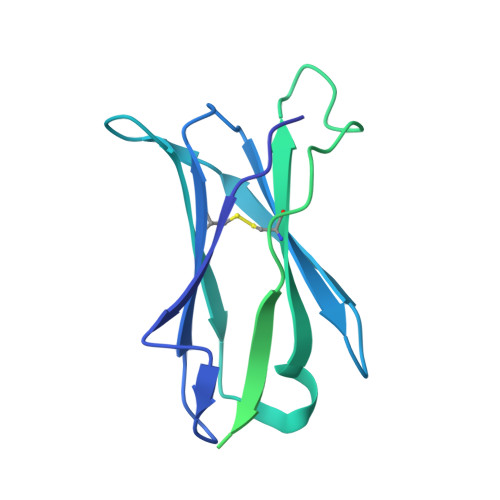
Structural basis for the activity and specificity of the immune checkpoint inhibitor lirilumab.

Lorig-Roach, N.Harpell, N.M.DuBois, R.M.

(2024) Sci Rep 14: 742-742

  • DOI: https://doi.org/10.1038/s41598-023-50262-6
  • Primary Citation Related Structures: 
    8TUI

  • PubMed Abstract: 

    The clinical success of immune checkpoint inhibitors has underscored the key role of the immune system in controlling cancer. Current FDA-approved immune checkpoint inhibitors target the regulatory receptor pathways of cytotoxic T-cells to enhance their anticancer responses. Despite an abundance of evidence that natural killer (NK) cells can also mediate potent anticancer activities, there are no FDA-approved inhibitors targeting NK cell specific checkpoint pathways. Lirilumab, the most clinically advanced NK cell checkpoint inhibitor, targets inhibitory killer immunoglobulin-like receptors (KIRs), however it has yet to conclusively demonstrate clinical efficacy. Here we describe the crystal structure of lirilumab in complex with the inhibitory KIR2DL3, revealing the precise epitope of lirilumab and the molecular mechanisms underlying KIR checkpoint blockade. Notably, the epitope includes several key amino acids that vary across the human population, and binding studies demonstrate the importance of these amino acids for lirilumab binding. These studies reveal how KIR variations in patients could influence the clinical efficacy of lirilumab and reveal general concepts for the development of immune checkpoint inhibitors targeting NK cells.


  • Organizational Affiliation
    • Department of Biomolecular Engineering, University of California Santa Cruz, Santa Cruz, CA, USA.

Macromolecule Content 

  • Total Structure Weight: 80.79 kDa 
  • Atom Count: 4,176 
  • Modeled Residue Count: 535 
  • Deposited Residue Count: 736 
  • Unique protein chains: 3

Macromolecules

Find similar proteins by:|  3D Structure
Entity ID: 1
MoleculeChains  Sequence LengthOrganismDetailsImage
Lirilumab Fab Heavy ChainA [auth H]265Homo sapiensMutation(s): 0 
Find similar proteins by:|  3D Structure
Entity ID: 2
MoleculeChains  Sequence LengthOrganismDetailsImage
Lirilumab Fab Light chainB [auth L]214Homo sapiensMutation(s): 0 
Find similar proteins by:|  3D Structure
Entity ID: 3
MoleculeChains  Sequence LengthOrganismDetailsImage
Killer cell immunoglobulin-like receptor 2DL3C [auth A]257Homo sapiensMutation(s): 0 
Gene Names: KIR2DL3CD158B2KIRCL23NKAT2
UniProt & NIH Common Fund Data Resources
Find proteins for P43628 (Homo sapiens)
Explore P43628 
Go to UniProtKB:  P43628
PHAROS:  P43628
GTEx:  ENSG00000243772 
Entity Groups
Sequence Clusters30% Identity50% Identity70% Identity90% Identity95% Identity100% Identity
UniProt GroupP43628
Glycosylation
Glycosylation Sites: 1Go to GlyGen: P43628-1
Sequence Annotations
Expand
Reference Sequence

Oligosaccharides

Help  
Entity ID: 4
MoleculeChains Length2D Diagram GlycosylationD Interactions
2-acetamido-2-deoxy-beta-D-glucopyranose-(1-2)-alpha-D-mannopyranose-(1-6)-[alpha-D-mannopyranose-(1-3)]beta-D-mannopyranose-(1-4)-2-acetamido-2-deoxy-beta-D-glucopyranose-(1-4)-[alpha-L-fucopyranose-(1-6)]2-acetamido-2-deoxy-beta-D-glucopyranoseD [auth B]7N-Glycosylation
Glycosylation Resources
GlyTouCan: G45889JQ
GlyCosmos: G45889JQ
GlyGen: G45889JQ

Experimental Data & Validation

Experimental Data

  • Method: X-RAY DIFFRACTION
  • Resolution: 2.75 Å
  • R-Value Free:  0.254 (Depositor), 0.253 (DCC) 
  • R-Value Work:  0.211 (Depositor), 0.210 (DCC) 
  • R-Value Observed: 0.213 (Depositor) 
Space Group: P 1 21 1
Unit Cell:
Length ( Å )Angle ( ˚ )
a = 58.379α = 90
b = 84.27β = 115.86
c = 62.268γ = 90
Software Package:
Software NamePurpose
PHENIXrefinement
PHENIXrefinement
HKL-2000data reduction
HKL-2000data scaling
PHENIXphasing

Structure Validation

View Full Validation Report


Entry History 

& Funding Information

Deposition Data


Funding OrganizationLocationGrant Number
Other privateUnited States--

Revision History  (Full details and data files)

  • Version 1.0: 2023-12-06
    Type: Initial release
  • Version 1.1: 2024-01-24
    Changes: Database references
  • Version 1.2: 2024-10-23
    Changes: Structure summary