8TT8 | pdb_00008tt8

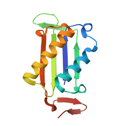
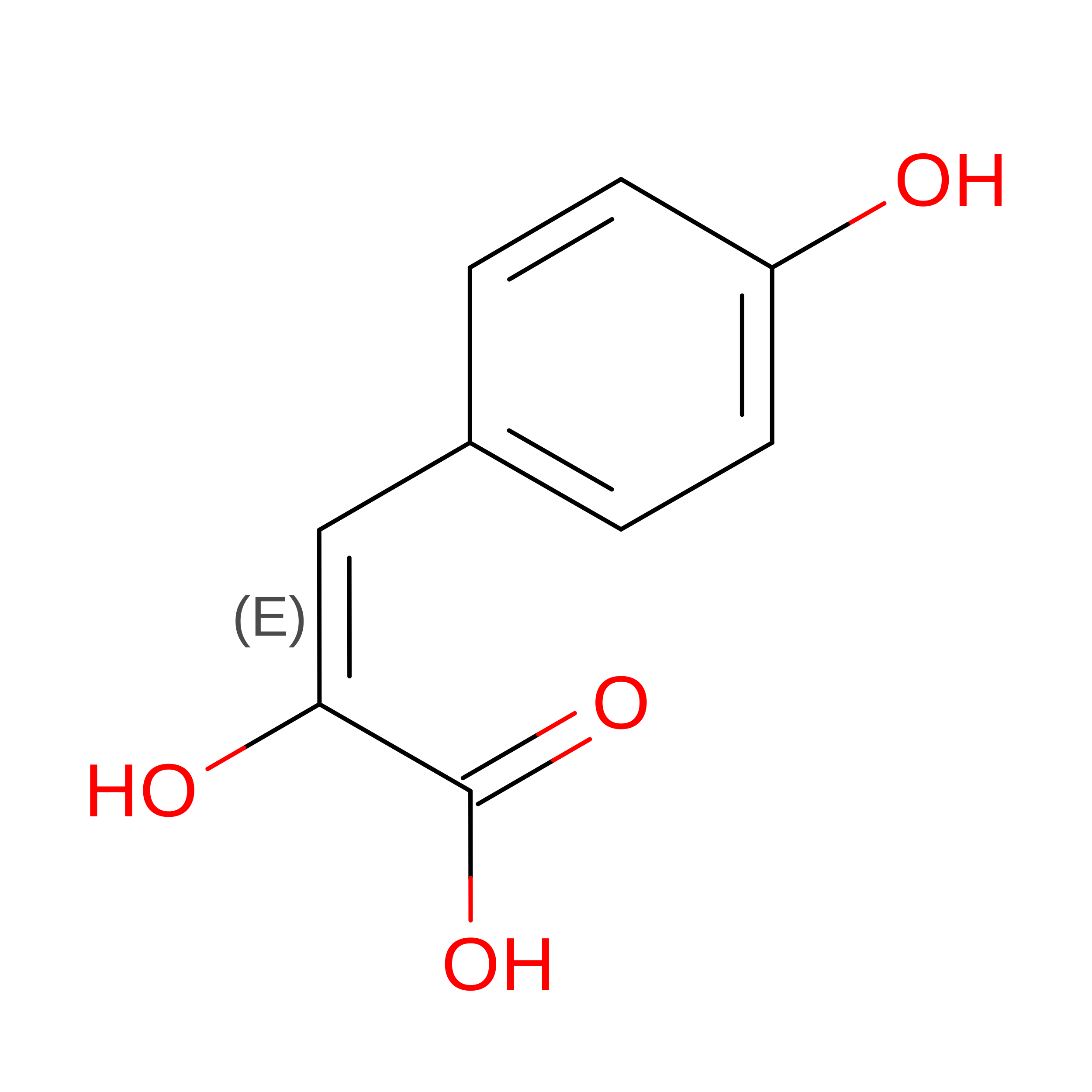
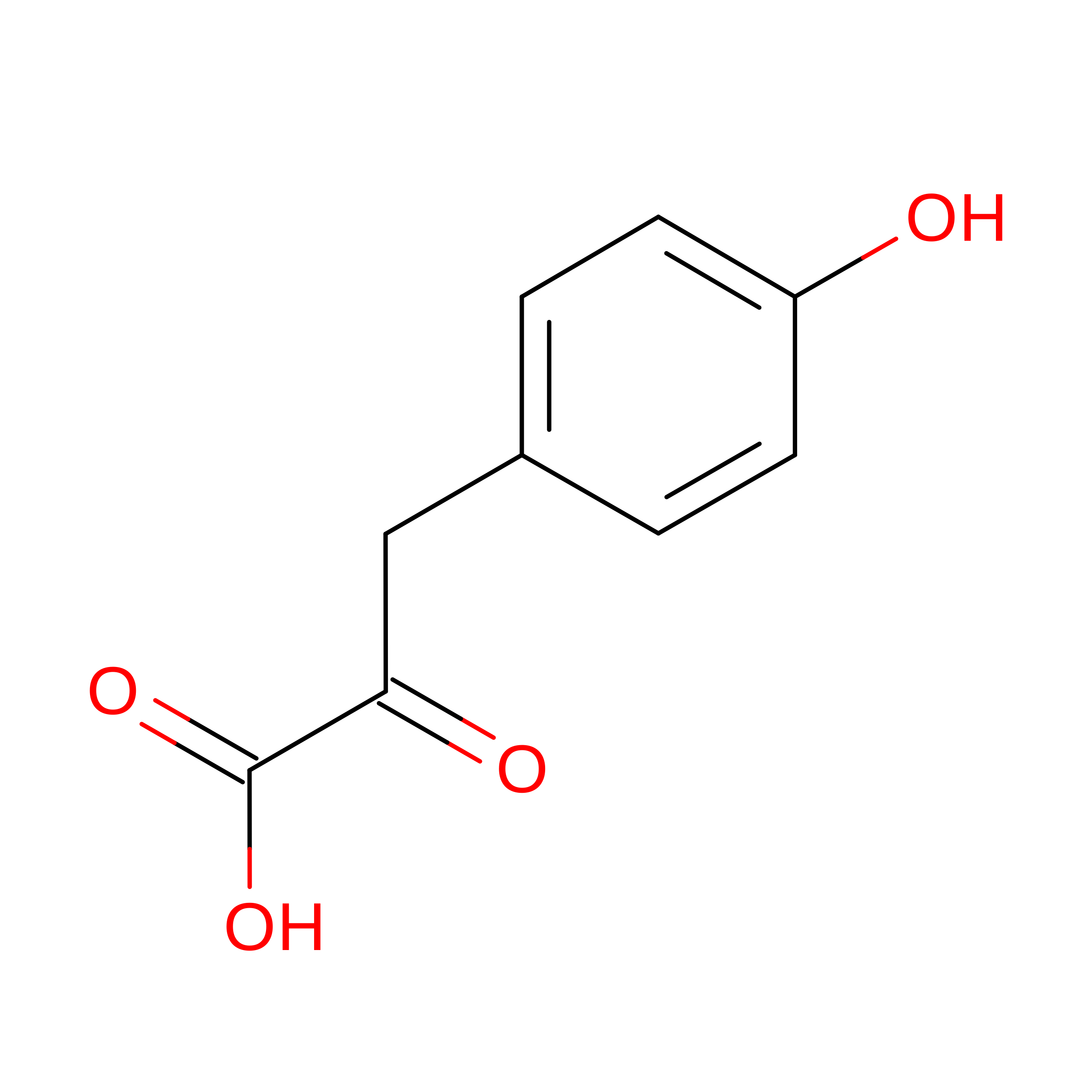
Joint Xray/Neutron structure of Macrophage Migration Inhibitory Factor (MIF) Bound to 4-hydroxyphenylpyruvate at room temperature


Experimental Data Snapshot

  • Method: X-RAY DIFFRACTION
  • Resolution: 1.75 Å
  • R-Value Free: 
    0.180 (Depositor), 0.183 (DCC) 
  • R-Value Work: 
    0.154 (Depositor), 0.155 (DCC) 
  • R-Value Observed: 
    0.155 (Depositor) 

  • Method: NEUTRON DIFFRACTION
  • Resolution: 2.53 Å
  • R-Value Free: 
    0.271 (Depositor) 
  • R-Value Work: 
    0.219 (Depositor) 
  • R-Value Observed: 
    0.221 (Depositor) 

Starting Model: experimental
View more details

wwPDB Validation 3D Report Full Report

Validation slider image for 8TT8

This is version 1.1 of the entry. See complete history. 

Literature

Identification of a key water molecule involved in the macrophage migration inhibitory factor-catalyzed tautomerization of para-hydroxyphenylpyruvate using neutron crystallography.

Schroder, G.C., Crichlow, G.V., Jablonowski, E., Pantouris, G., Nix, J., Chayen, N., Meilleur, F., Lolis, E.J.

(2026) Protein Sci 35: e70666-e70666

  • DOI: https://doi.org/10.1002/pro.70666
  • Primary Citation Related Structures: 
    8TT8, 8TT9

  • PubMed Abstract: 

    Neutron crystallography was used to determine a 2.5-Å resolution all-atom structure of macrophage migration inhibitory factor (MIF) interacting with 3-(4-hydroxyphenyl)-pyruvate (HPP). MIF is a pro-inflammatory, pro-tumorigenic protein that may be an attractive therapeutic target. MIF catalyzes the interconversion of the keto and enol forms of HPP by a tautomerase reaction. Although HPP is evidently not a physiological substrate of MIF, many compounds that inhibit this activity in enzymatic assays have been found also to inhibit physiological activities of MIF. Therefore, the MIF-catalyzed HPP tautomerization reaction is used in initial screening of compounds in the search for inhibitors of MIF physiological activity. The neutron diffraction-derived crystal structure reveals the position of a water molecule involved in the tautomerization reaction, and also confirms the charged state of lysine-32 in the active site. The structure confirms the previously proposed catalytic mechanism of MIF, with the N-terminal Pro-1 abstracting a proton to generate an HPP enolate intermediate which is subsequently protonated. The structure reported herein reveals that this proton is supplied by a neighboring water molecule. Along with the neutron structure, a room-temperature synchrotron x-ray crystal structure reveals a covalent adduct between HPP and MIF. While this adduct is a result of radiation-induced chemistry, its formation confirms the catalytic role of the active site residue because a covalent complex could only form if the reactive carbon of the substrate is correctly positioned by the enzyme.


  • Organizational Affiliation: 
    • Department of Molecular and Structural Biochemistry, North Carolina State University, Raleigh, North Carolina, USA.

Macromolecule Content 

  • Total Structure Weight: 37.67 kDa 
  • Atom Count: 3,043 
  • Modeled Residue Count: 342 
  • Deposited Residue Count: 342 
  • Unique protein chains: 1

Macromolecules

Find similar proteins by:|  3D Structure
Entity ID: 1
MoleculeChains  Sequence LengthOrganismDetailsImage
Macrophage migration inhibitory factor
A, B, C
114Homo sapiensMutation(s): 0 
Gene Names: MIF, GLIF, MMIF
EC: 5.3.2.1 (PDB Primary Data), 5.3.3.12 (PDB Primary Data)
UniProt & NIH Common Fund Data Resources
Find proteins for P14174 (Homo sapiens)
Explore P14174 
Go to UniProtKB:  P14174
PHAROS:  P14174
GTEx:  ENSG00000240972 
Entity Groups
Sequence Clusters30% Identity50% Identity70% Identity90% Identity95% Identity100% Identity
UniProt GroupP14174
Sequence Annotations
Expand
Reference Sequence

Experimental Data & Validation

Experimental Data

  • Method: X-RAY DIFFRACTION
  • Resolution: 1.75 Å
  • R-Value Free:  0.180 (Depositor), 0.183 (DCC) 
  • R-Value Work:  0.154 (Depositor), 0.155 (DCC) 
  • R-Value Observed: 0.155 (Depositor) 
  • Method: NEUTRON DIFFRACTION
  • Resolution: 2.53 Å
  • R-Value Free:  0.271 (Depositor) 
  • R-Value Work:  0.219 (Depositor) 
  • R-Value Observed: 0.221 (Depositor) 
Space Group: P 21 21 21
Unit Cell:
Length ( Å )Angle ( ˚ )
a = 68.613α = 90
b = 69.231β = 90
c = 89.185γ = 90
Software Package:
Software NamePurpose
PHENIXrefinement

Structure Validation

View Full Validation Report


Entry History 

& Funding Information

Deposition Data


Funding OrganizationLocationGrant Number
Department of Energy (DOE, United States)United States--

Revision History  (Full details and data files)

  • Version 1.0: 2025-08-27
    Type: Initial release
  • Version 1.1: 2026-09-09
    Changes: Database references