Characterisation of the heme aqua-ligand coordination environment in an engineered peroxygenase cytochrome P450 variant.
Podgorski, M.N., Lee, J.H.Z., Harbort, J.S., Nguyen, G.T.H., Doherty, D.Z., Donald, W.A., Harmer, J.R., Bruning, J.B., Bell, S.G.(2023) J Inorg Biochem 249: 112391-112391
- PubMed: 37837941 Search on PubMed
- DOI: https://doi.org/10.1016/j.jinorgbio.2023.112391
- Primary Citation Related Structures:
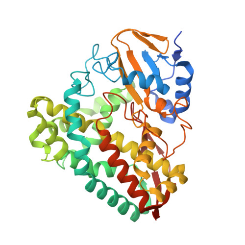
8TAW, 8TAY, 8TNK - PubMed Abstract:
The cytochrome P450 enzymes (CYPs) are heme-thiolate monooxygenases that catalyse the insertion of an oxygen atom into the C-H bonds of organic molecules. In most CYPs, the activation of dioxygen by the heme is aided by an acid-alcohol pair of residues located in the I-helix of the enzyme. Mutation of the threonine residue of this acid-alcohol pair of CYP199A4, from the bacterium Rhodospeudomonas palustris HaA2, to a glutamate residue induces peroxygenase activity. In the X-ray crystal structures of this variant an interaction of the glutamate side chain and the distal aqua ligand of the heme was observed and this results in this ligand not being readily displaced in the peroxygenase mutant on the addition of substrate. Here we use a range of bulky hydrophobic and nitrogen donor containing ligands in an attempt to displace the distal aqua ligand of the T252E mutant of CYP199A4. Ligand binding was assessed by UV-visible absorbance spectroscopy, native mass spectrometry, electron paramagnetic resonance and X-ray crystallography. None of the ligands tested, even the nitrogen donor ligands which bind directly to the iron in the wild-type enzyme, resulted in displacement of the aqua ligand. Therefore, modification of the I-helix threonine residue to a glutamate residue results in a significant strengthening of the ferric distal aqua ligand. This ligand was not displaced using any of the ligands during this study and this provides a rationale as to why this mutant can shutdown the monooxygenase pathway of this enzyme and switch to peroxygenase activity.
- Department of Chemistry, University of Adelaide, Adelaide, SA 5005, Australia.
Organizational Affiliation: