8TGT | pdb_00008tgt

Structure of human C4b-binding protein alpha chain CCP domains 1 and 2 in complex with the hypervariable region of group A Streptococcus M68 protein


Experimental Data Snapshot

  • Method: X-RAY DIFFRACTION
  • Resolution: 2.50 Å
  • R-Value Free: 
    0.344 (Depositor), 0.340 (DCC) 
  • R-Value Work: 
    0.313 (Depositor), 0.310 (DCC) 
  • R-Value Observed: 
    0.316 (Depositor) 

wwPDB Validation   3D Report Full Report


Ligand Structure Quality Assessment 


This is version 1.1 of the entry. See complete history


Literature

Conservation of C4BP-binding sequence patterns in Streptococcus pyogenes M and Enn proteins.

Kolesinski, P.McGowan, M.Botteaux, A.Smeesters, P.R.Ghosh, P.

(2024) J Biological Chem 300: 107478-107478

  • DOI: https://doi.org/10.1016/j.jbc.2024.107478
  • Primary Citation of Related Structures:  
    8TCB, 8TGT

  • PubMed Abstract: 

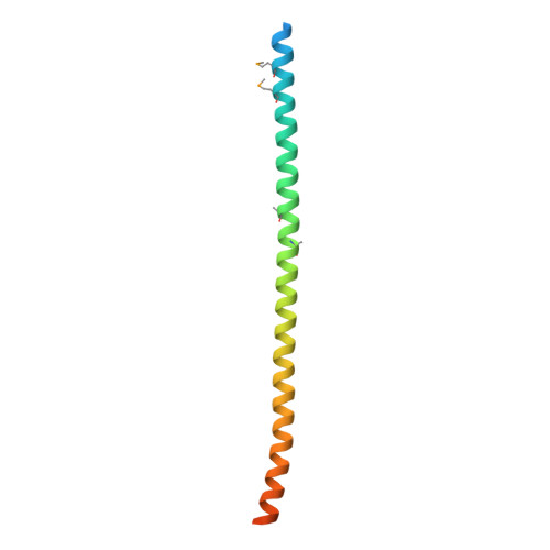
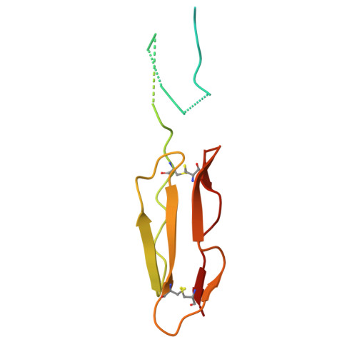
    Antigenically sequence variable M proteins of the major bacterial pathogen Streptococcus pyogenes (Strep A) are responsible for recruiting human C4b-binding protein (C4BP) to the bacterial surface, which enables Strep A to evade destruction by the immune system. The most sequence divergent portion of M proteins, the hypervariable region (HVR), is responsible for binding C4BP. Structural evidence points to the conservation of two C4BP-binding sequence patterns (M2 and M22) in the HVR of numerous M proteins, with this conservation applicable to vaccine immunogen design. These two patterns, however, only partially explain C4BP binding by Strep A. Here, we identified several M proteins that lack these patterns but still bind C4BP and determined the structures of two, M68 and M87 HVRs, in complex with a C4BP fragment. Mutagenesis of these M proteins led to the identification of amino acids that are crucial for C4BP binding, enabling formulation of new C4BP-binding patterns. Mutagenesis was also carried out on M2 and M22 proteins to refine or generate experimentally grounded C4BP-binding patterns. The M22 pattern was the most prevalent among M proteins, followed by the M87 and M2 patterns, while the M68 pattern was rare. These patterns, except for M68, were also evident in numerous M-like Enn proteins. Binding of C4BP via these patterns to Enn proteins was verified. We conclude that C4BP-binding patterns occur frequently in Strep A strains of differing M types, being present in their M or Enn proteins, or frequently both, providing further impetus for their use as vaccine immunogens.


  • Organizational Affiliation
    • Department of Chemistry and Biochemistry, University of California, San Diego La Jolla, California, USA.

Macromolecules
Find similar proteins by:  (by identity cutoff)  |  3D Structure
Entity ID: 1
MoleculeChains Sequence LengthOrganismDetailsImage
M protein
A, B
104Streptococcus pyogenesMutation(s): 0 
Gene Names: emm
UniProt
Find proteins for A0A059T9V1 (Streptococcus pyogenes)
Explore A0A059T9V1 
Go to UniProtKB:  A0A059T9V1
Entity Groups  
Sequence Clusters30% Identity50% Identity70% Identity90% Identity95% Identity100% Identity
UniProt GroupA0A059T9V1
Sequence Annotations
Expand
  • Reference Sequence
Find similar proteins by:  (by identity cutoff)  |  3D Structure
Entity ID: 2
MoleculeChains Sequence LengthOrganismDetailsImage
C4b-binding protein alpha chain128Homo sapiensMutation(s): 0 
Gene Names: C4BPAC4BP
UniProt & NIH Common Fund Data Resources
Find proteins for P04003 (Homo sapiens)
Explore P04003 
Go to UniProtKB:  P04003
PHAROS:  P04003
GTEx:  ENSG00000123838 
Entity Groups  
Sequence Clusters30% Identity50% Identity70% Identity90% Identity95% Identity100% Identity
UniProt GroupP04003
Sequence Annotations
Expand
  • Reference Sequence
Experimental Data & Validation

Experimental Data

  • Method: X-RAY DIFFRACTION
  • Resolution: 2.50 Å
  • R-Value Free:  0.344 (Depositor), 0.340 (DCC) 
  • R-Value Work:  0.313 (Depositor), 0.310 (DCC) 
  • R-Value Observed: 0.316 (Depositor) 
Space Group: P 21 21 2
Unit Cell:
Length ( Å )Angle ( ˚ )
a = 107.037α = 90
b = 49.664β = 90
c = 80.403γ = 90
Software Package:
Software NamePurpose
PHENIXrefinement
XDSdata reduction
pointlessdata scaling
Aimlessdata scaling
AutoSolphasing

Structure Validation

View Full Validation Report


Ligand Structure Quality Assessment 


Entry History & Funding Information

Deposition Data


Funding OrganizationLocationGrant Number
National Institutes of Health/National Institute Of Allergy and Infectious Diseases (NIH/NIAID)United StatesNIH R01 AI154149

Revision History  (Full details and data files)

  • Version 1.0: 2024-07-17
    Type: Initial release
  • Version 1.1: 2024-11-20
    Changes: Database references, Structure summary