8TFH

Ricin in complex with Fab JB4


Experimental Data Snapshot

  • Method: X-RAY DIFFRACTION
  • Resolution: 3.29 Å
  • R-Value Free: 0.334 
  • R-Value Work: 0.274 
  • R-Value Observed: 0.277 

wwPDB Validation   3D Report Full Report


This is version 1.0 of the entry. See complete history


Literature

Structural Basis of Antibody-Mediated Inhibition of Ricin Toxin Attachment to Host Cells.

Vance, D.J.Rudolph, M.J.Davis, S.A.Mantis, N.J.

(2023) Biochemistry 62: 3181-3187

  • DOI: https://doi.org/10.1021/acs.biochem.3c00480
  • Primary Citation of Related Structures:  
    8TFH, 8TFL

  • PubMed Abstract: 

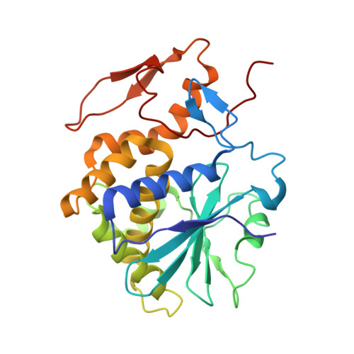
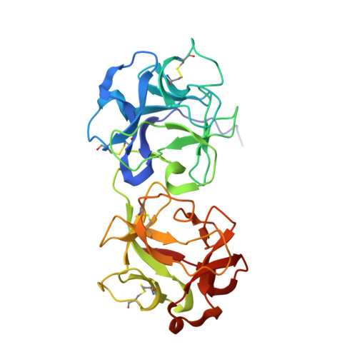
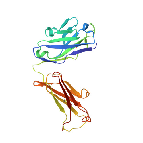
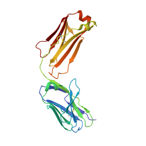
    Monoclonal antibodies, JB4 and SylH3, neutralize ricin toxin (RT) by inhibiting the galactose-specific lectin activity of the B subunit of the toxin (RTB), which is required for cell attachment and entry. It is not immediately apparent how the antibodies accomplish this feat, considering that RTB consists of two globular domains (D1, D2) each divided into three homologous subdomains (α, β, γ) with the two functional galactosyl-specific carbohydrate recognition domains (CRDs) situated on opposite poles (subdomains 1α and 2γ). Here, we report the X-ray crystal structures of JB4 and SylH3 Fab fragments bound to RTB in the context of RT. The structures revealed that neither Fab obstructed nor induced detectable conformational alterations in subdomains 1α or 2γ. Rather, JB4 and SylH3 Fabs recognize nearly identical epitopes within an ancillary carbohydrate recognition pocket located in subdomain 1β. Despite limited amino acid sequence similarity between SylH3 and JB4 Fabs, each paratope inserts a Phe side chain from the heavy (H) chain complementarity determining region (CDR3) into the 1β CRD pocket, resulting in local aromatic stacking interactions that potentially mimic a ligand interaction. Reconciling the fact that stoichiometric amounts of SylH3 and JB4 are sufficient to disarm RTB's lectin activity without evidence of allostery, we propose that subdomain 1β functions as a "coreceptor" required to stabilize glycan interactions principally mediated by subdomains 1α and 2γ. Further investigation into subdomain 1β will yield fundamental insights into the large family of R-type lectins and open novel avenues for countermeasures aimed at preventing toxin uptake into vulnerable tissues and cells.


  • Organizational Affiliation

    Division of Infectious Diseases, Wadsworth Center, New York State Department of Health, Albany, New York 12208, United States.


Macromolecules
Find similar proteins by:  (by identity cutoff)  |  3D Structure
Entity ID: 1
MoleculeChains Sequence LengthOrganismDetailsImage
Ricin A chain260Ricinus communisMutation(s): 0 
EC: 3.2.2.22
UniProt
Find proteins for P02879 (Ricinus communis)
Explore P02879 
Go to UniProtKB:  P02879
Entity Groups  
Sequence Clusters30% Identity50% Identity70% Identity90% Identity95% Identity100% Identity
UniProt GroupP02879
Sequence Annotations
Expand
  • Reference Sequence
Find similar proteins by:  (by identity cutoff)  |  3D Structure
Entity ID: 2
MoleculeChains Sequence LengthOrganismDetailsImage
Ricin B chain262Ricinus communisMutation(s): 0 
EC: 3.2.2.22
UniProt
Find proteins for P02879 (Ricinus communis)
Explore P02879 
Go to UniProtKB:  P02879
Entity Groups  
Sequence Clusters30% Identity50% Identity70% Identity90% Identity95% Identity100% Identity
UniProt GroupP02879
Sequence Annotations
Expand
  • Reference Sequence
Find similar proteins by:  (by identity cutoff)  |  3D Structure
Entity ID: 3
MoleculeChains Sequence LengthOrganismDetailsImage
JB4 monoclonal antibody heavy chainC [auth H]223Mus musculusMutation(s): 0 
Entity Groups  
Sequence Clusters30% Identity50% Identity70% Identity90% Identity95% Identity100% Identity
Sequence Annotations
Expand
  • Reference Sequence
Find similar proteins by:  (by identity cutoff)  |  3D Structure
Entity ID: 4
MoleculeChains Sequence LengthOrganismDetailsImage
JB4 monoclonal antibody light chainD [auth L]210Mus musculusMutation(s): 0 
Entity Groups  
Sequence Clusters30% Identity50% Identity70% Identity90% Identity95% Identity100% Identity
Sequence Annotations
Expand
  • Reference Sequence
Oligosaccharides

Help

Entity ID: 5
MoleculeChains Length2D Diagram Glycosylation3D Interactions
alpha-L-fucopyranose-(1-3)-[2-acetamido-2-deoxy-beta-D-glucopyranose-(1-4)]2-acetamido-2-deoxy-beta-D-glucopyranoseE [auth C]3N-Glycosylation
Glycosylation Resources
GlyTouCan:  G11971MR
GlyCosmos:  G11971MR
GlyGen:  G11971MR
Entity ID: 6
MoleculeChains Length2D Diagram Glycosylation3D Interactions
beta-D-mannopyranose-(1-4)-2-acetamido-2-deoxy-beta-D-glucopyranose-(1-4)-2-acetamido-2-deoxy-beta-D-glucopyranoseF [auth D],
G [auth E]
3N-Glycosylation
Glycosylation Resources
GlyTouCan:  G15407YE
GlyCosmos:  G15407YE
GlyGen:  G15407YE
Experimental Data & Validation

Experimental Data

  • Method: X-RAY DIFFRACTION
  • Resolution: 3.29 Å
  • R-Value Free: 0.334 
  • R-Value Work: 0.274 
  • R-Value Observed: 0.277 
  • Space Group: I 41 2 2
Unit Cell:
Length ( Å )Angle ( ˚ )
a = 139.358α = 90
b = 139.358β = 90
c = 444.613γ = 90
Software Package:
Software NamePurpose
PHENIXrefinement
HKL-2000data reduction
HKL-2000data scaling
PHASERphasing

Structure Validation

View Full Validation Report


Entry History & Funding Information

Deposition Data


Funding OrganizationLocationGrant Number
National Institutes of Health/National Institute Of Allergy and Infectious Diseases (NIH/NIAID)United StatesHHSN272201400021C

Revision History  (Full details and data files)

  • Version 1.0: 2024-04-17
    Type: Initial release