In Situ Structural Observation of a Substrate- and Peroxide-Bound High-Spin Ferric-Hydroperoxo Intermediate in the P450 Enzyme CYP121.
Nguyen, R.C., Davis, I., Dasgupta, M., Wang, Y., Simon, P.S., Butryn, A., Makita, H., Bogacz, I., Dornevil, K., Aller, P., Bhowmick, A., Chatterjee, R., Kim, I.S., Zhou, T., Mendez, D., Paley, D.W., Fuller, F., Alonso Mori, R., Batyuk, A., Sauter, N.K., Brewster, A.S., Orville, A.M., Yachandra, V.K., Yano, J., Kern, J.F., Liu, A.(2023) J Am Chem Soc 145: 25120-25133
- PubMed: 37939223 Search on PubMedSearch on PubMed Central
- DOI: https://doi.org/10.1021/jacs.3c04991
- Primary Citation Related Structures:
8TDP, 8TDQ - PubMed Abstract:
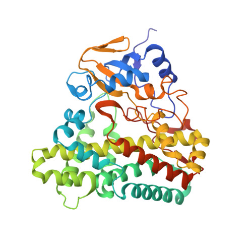
The P450 enzyme CYP121 from Mycobacterium tuberculosis catalyzes a carbon-carbon (C-C) bond coupling cyclization of the dityrosine substrate containing a diketopiperazine ring, cyclo (l-tyrosine-l-tyrosine) (cYY). An unusual high-spin ( S = 5/2) ferric intermediate maximizes its population in less than 5 ms in the rapid freeze-quenching study of CYP121 during the shunt reaction with peracetic acid or hydrogen peroxide in acetic acid solution. We show that this intermediate can also be observed in the crystalline state by EPR spectroscopy. By developing an on-demand-rapid-mixing method for time-resolved serial femtosecond crystallography with X-ray free-electron laser (tr-SFX-XFEL) technology covering the millisecond time domain and without freezing, we structurally monitored the reaction in situ at room temperature. After a 200 ms peracetic acid reaction with the cocrystallized enzyme-substrate microcrystal slurry, a ferric-hydroperoxo intermediate is observed, and its structure is determined at 1.85 Å resolution. The structure shows a hydroperoxyl ligand between the heme and the native substrate, cYY. The oxygen atoms of the hydroperoxo are 2.5 and 3.2 Å from the iron ion. The end-on binding ligand adopts a near-side-on geometry and is weakly associated with the iron ion, causing the unusual high-spin state. This compound 0 intermediate, spectroscopically and structurally observed during the catalytic shunt pathway, reveals a unique binding mode that deviates from the end-on compound 0 intermediates in other heme enzymes. The hydroperoxyl ligand is only 2.9 Å from the bound cYY, suggesting an active oxidant role of the intermediate for direct substrate oxidation in the nonhydroxylation C-C bond coupling chemistry.
- Department of Chemistry, University of Texas, San Antonio, Texas 78249, United States.
Organizational Affiliation: