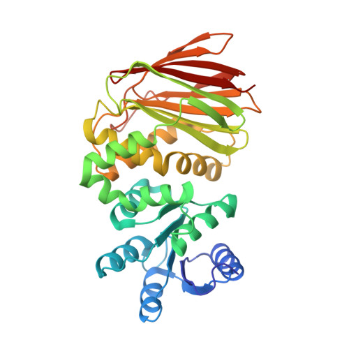
Structure of phosphorylated-like RssB, the adaptor delivering sigma s to the ClpXP proteolytic machinery, reveals an interface switch for activation.
Brugger, C., Schwartz, J., Novick, S., Tong, S., Hoskins, J.R., Majdalani, N., Kim, R., Filipovski, M., Wickner, S., Gottesman, S., Griffin, P.R., Deaconescu, A.M.(2023) J Biological Chem 299: 105440-105440
- PubMed: 37949227 Search on PubMedSearch on PubMed Central
- DOI: https://doi.org/10.1016/j.jbc.2023.105440
- Primary Citation Related Structures:
8T85 - PubMed Abstract:
In enterobacteria such as Escherichia coli, the general stress response is mediated by σ s , the stationary phase dissociable promoter specificity subunit of RNA polymerase. σ s is degraded by ClpXP during active growth in a process dependent on the RssB adaptor, which is thought to be stimulated by the phosphorylation of a conserved aspartate in its N-terminal receiver domain. Here we present the crystal structure of full-length RssB bound to a beryllofluoride phosphomimic. Compared to the structure of RssB bound to the IraD anti-adaptor, our new RssB structure with bound beryllofluoride reveals conformational differences and coil-to-helix transitions in the C-terminal region of the RssB receiver domain and in the interdomain segmented helical linker. These are accompanied by masking of the α4-β5-α5 (4-5-5) "signaling" face of the RssB receiver domain by its C-terminal domain. Critically, using hydrogen-deuterium exchange mass spectrometry, we identify σ s -binding determinants on the 4-5-5 face, implying that this surface needs to be unmasked to effect an interdomain interface switch and enable full σ s engagement and hand-off to ClpXP. In activated receiver domains, the 4-5-5 face is often the locus of intermolecular interactions, but its masking by intramolecular contacts upon phosphorylation is unusual, emphasizing that RssB is a response regulator that undergoes atypical regulation.
- Laboratories of Molecular Medicine, Department of Molecular Biology, Cell Biology and Biochemistry, Brown University, Providence, Rhode Island, USA.
Organizational Affiliation: