8T3W

Structure of Bre1-nucleosome complex - state2


Experimental Data Snapshot

  • Method: ELECTRON MICROSCOPY
  • Resolution: 3.25 Å
  • Aggregation State: PARTICLE 
  • Reconstruction Method: SINGLE PARTICLE 

wwPDB Validation   3D Report Full Report


This is version 1.3 of the entry. See complete history


Literature

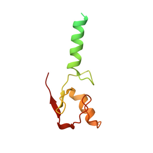
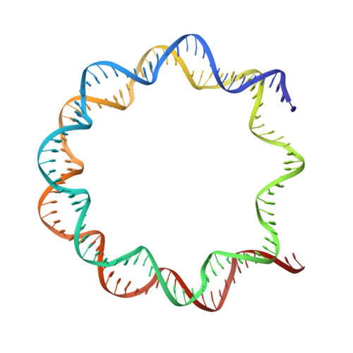
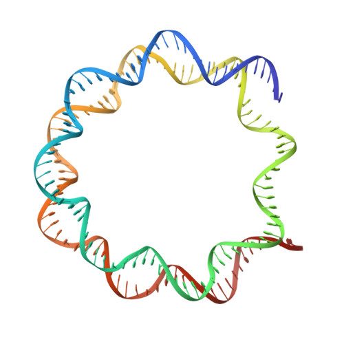
Mechanism of histone H2B monoubiquitination by Bre1.

Zhao, F.Hicks, C.W.Wolberger, C.

(2023) Nat Struct Mol Biol 30: 1623-1627

  • DOI: https://doi.org/10.1038/s41594-023-01137-x
  • Primary Citation of Related Structures:  
    8T3T, 8T3W, 8T3Y

  • PubMed Abstract: 

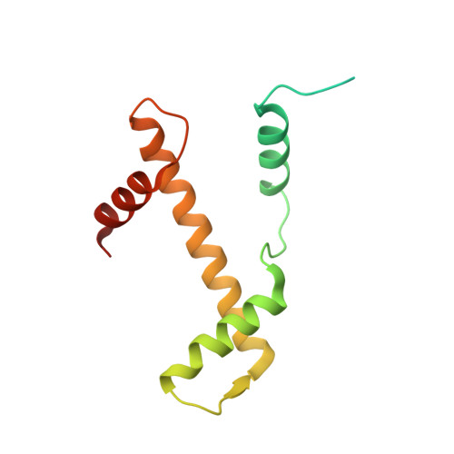
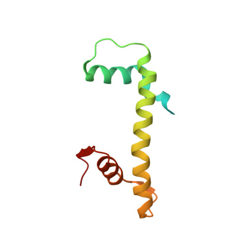
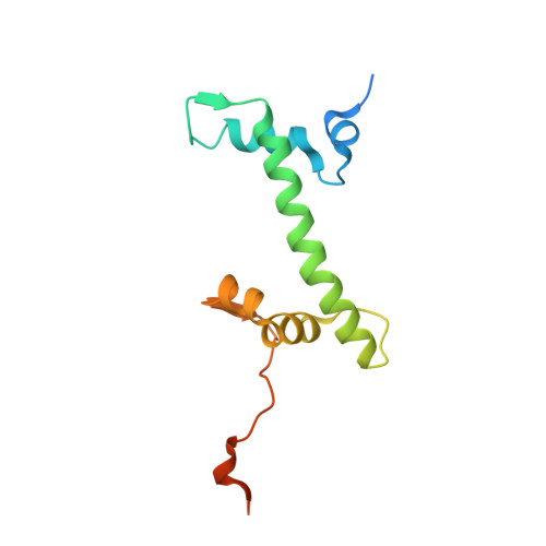
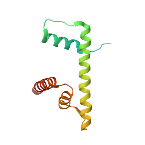
    Monoubiquitination of histone H2B-K120/123 plays several roles in regulating transcription, DNA replication and the DNA damage response. The structure of a nucleosome in complex with the dimeric RING E3 ligase Bre1 reveals that one RING domain binds to the nucleosome acidic patch, where it can position the E2 ubiquitin conjugating enzyme Rad6, while the other RING domain contacts the DNA. Comparisons with H2A-specific E3 ligases suggest a general mechanism of tuning histone specificity via the non-E2-binding RING domain.


  • Organizational Affiliation

    Department of Biophysics and Biophysical Chemistry, Johns Hopkins University School of Medicine, Baltimore, MD, USA.


Macromolecules

Find similar proteins by:  (by identity cutoff)  |  3D Structure
Entity ID: 1
MoleculeChains Sequence LengthOrganismDetailsImage
Histone H3.2
A, E
135Xenopus laevisMutation(s): 1 
UniProt
Find proteins for P84233 (Xenopus laevis)
Explore P84233 
Go to UniProtKB:  P84233
Entity Groups  
Sequence Clusters30% Identity50% Identity70% Identity90% Identity95% Identity100% Identity
UniProt GroupP84233
Sequence Annotations
Expand
  • Reference Sequence
Find similar proteins by:  (by identity cutoff)  |  3D Structure
Entity ID: 2
MoleculeChains Sequence LengthOrganismDetailsImage
Histone H4
B, F
102Xenopus laevisMutation(s): 0 
UniProt
Find proteins for P62799 (Xenopus laevis)
Explore P62799 
Go to UniProtKB:  P62799
Entity Groups  
Sequence Clusters30% Identity50% Identity70% Identity90% Identity95% Identity100% Identity
UniProt GroupP62799
Sequence Annotations
Expand
  • Reference Sequence
Find similar proteins by:  (by identity cutoff)  |  3D Structure
Entity ID: 3
MoleculeChains Sequence LengthOrganismDetailsImage
Histone H2A type 1
C, G
129Xenopus laevisMutation(s): 0 
UniProt
Find proteins for P06897 (Xenopus laevis)
Explore P06897 
Go to UniProtKB:  P06897
Entity Groups  
Sequence Clusters30% Identity50% Identity70% Identity90% Identity95% Identity100% Identity
UniProt GroupP06897
Sequence Annotations
Expand
  • Reference Sequence
Find similar proteins by:  (by identity cutoff)  |  3D Structure
Entity ID: 4
MoleculeChains Sequence LengthOrganismDetailsImage
Histone H2B
D, H
122Xenopus laevisMutation(s): 1 
UniProt
Find proteins for P02281 (Xenopus laevis)
Explore P02281 
Go to UniProtKB:  P02281
Entity Groups  
Sequence Clusters30% Identity50% Identity70% Identity90% Identity95% Identity100% Identity
UniProt GroupP02281
Sequence Annotations
Expand
  • Reference Sequence
Find similar proteins by:  (by identity cutoff)  |  3D Structure
Entity ID: 7
MoleculeChains Sequence LengthOrganismDetailsImage
E3 ubiquitin-protein ligase BRE1
K, L
110Saccharomyces cerevisiaeMutation(s): 0 
Gene Names: BRE1YDL074C
EC: 2.3.2.27
UniProt
Find proteins for Q07457 (Saccharomyces cerevisiae (strain ATCC 204508 / S288c))
Explore Q07457 
Go to UniProtKB:  Q07457
Entity Groups  
Sequence Clusters30% Identity50% Identity70% Identity90% Identity95% Identity100% Identity
UniProt GroupQ07457
Sequence Annotations
Expand
  • Reference Sequence
Find similar nucleic acids by:  (by identity cutoff)  |  3D Structure
Entity ID: 5
MoleculeChains LengthOrganismImage
601 DNA strand 1146synthetic construct
Sequence Annotations
Expand
  • Reference Sequence
Find similar nucleic acids by:  (by identity cutoff)  |  3D Structure
Entity ID: 6
MoleculeChains LengthOrganismImage
601 DNA strand 2146synthetic construct
Sequence Annotations
Expand
  • Reference Sequence
Experimental Data & Validation

Experimental Data

  • Method: ELECTRON MICROSCOPY
  • Resolution: 3.25 Å
  • Aggregation State: PARTICLE 
  • Reconstruction Method: SINGLE PARTICLE 
EM Software:
TaskSoftware PackageVersion
MODEL REFINEMENTPHENIX1.20.1
MODEL REFINEMENTCoot0.8.9.2

Structure Validation

View Full Validation Report


Entry History & Funding Information

Deposition Data


Funding OrganizationLocationGrant Number
National Institutes of Health/National Institute of General Medical Sciences (NIH/NIGMS)United StatesGM130393

Revision History  (Full details and data files)

  • Version 1.0: 2023-10-18
    Type: Initial release
  • Version 1.1: 2023-11-01
    Changes: Database references
  • Version 1.2: 2023-11-08
    Changes: Database references
  • Version 1.3: 2023-11-22
    Changes: Database references