8SRX

Crystal structure of BAK-BAX heterodimer with lysoPC


Experimental Data Snapshot

  • Method: X-RAY DIFFRACTION
  • Resolution: 2.09 Å
  • R-Value Free: 0.265 
  • R-Value Work: 0.216 
  • R-Value Observed: 0.221 

wwPDB Validation   3D Report Full Report


Ligand Structure Quality Assessment 


This is version 1.0 of the entry. See complete history


Literature

Sequence differences between BAX and BAK core domains manifest as differences in their interactions with lipids.

Miller, M.S.Cowan, A.D.Brouwer, J.M.Smyth, S.T.Peng, L.Wardak, A.Z.Uren, R.T.Luo, C.Roy, M.J.Shah, S.Tan, Z.Reid, G.E.Colman, P.M.Czabotar, P.E.

(2023) FEBS J 

  • DOI: https://doi.org/10.1111/febs.17031
  • Primary Citation of Related Structures:  
    8G1T, 8SPE, 8SPF, 8SPZ, 8SRX, 8SRY, 8SVK

  • PubMed Abstract: 

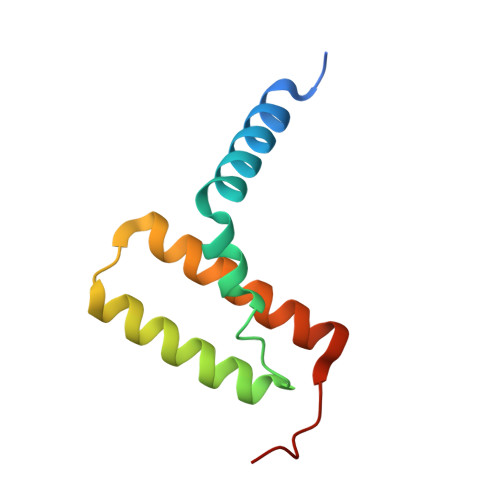
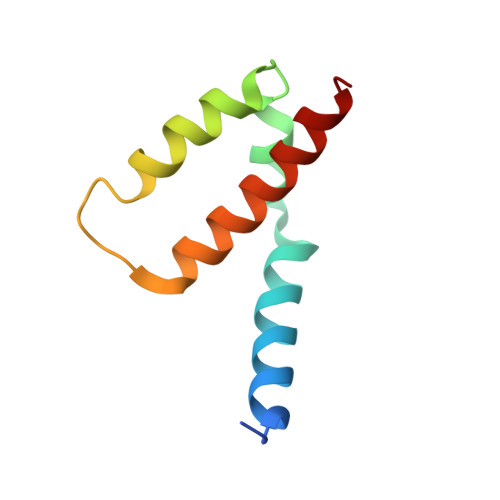
    The B-cell lymphoma 2 (BCL2) family members, BCL2-associated protein X (BAX) and BCL2 homologous antagonist killer (BAK), are required for programmed cell death via the mitochondrial pathway. When cells are stressed, damaged or redundant, the balance of power between the BCL2 family of proteins shifts towards BAX and BAK, allowing their transition from an inactive, monomeric state to a membrane-active oligomeric form that releases cytochrome c from the mitochondrial intermembrane space. That oligomeric state has an essential intermediate, a symmetric homodimer of BAX or BAK. Here we describe crystal structures of dimers of the core domain of BAX, comprising its helices α2-α5. These structures provide an atomic resolution description of the interactions that drive BAX homo-dimerisation and insights into potential interaction between core domain dimers and membrane lipids. The previously identified BAK lipid-interacting sites are not conserved with BAX and are likely to determine the differences between them in their interactions with lipids. We also describe structures of heterodimers of BAK/BAX core domains, yielding further insight into the differences in lipid binding between BAX and BAK.


  • Organizational Affiliation

    Walter and Eliza Hall Institute of Medical Research, Parkville, Vic., Australia.


Macromolecules
Find similar proteins by:  (by identity cutoff)  |  3D Structure
Entity ID: 1
MoleculeChains Sequence LengthOrganismDetailsImage
Bcl-2 homologous antagonist/killer
A, C
91Homo sapiensMutation(s): 0 
Gene Names: BAK1BAKBCL2L7CDN1
UniProt & NIH Common Fund Data Resources
Find proteins for Q16611 (Homo sapiens)
Explore Q16611 
Go to UniProtKB:  Q16611
PHAROS:  Q16611
GTEx:  ENSG00000030110 
Entity Groups  
Sequence Clusters30% Identity50% Identity70% Identity90% Identity95% Identity100% Identity
UniProt GroupQ16611
Sequence Annotations
Expand
  • Reference Sequence
Find similar proteins by:  (by identity cutoff)  |  3D Structure
Entity ID: 2
MoleculeChains Sequence LengthOrganismDetailsImage
Apoptosis regulator BAX
B, D
80Homo sapiensMutation(s): 0 
Gene Names: BAXBCL2L4
UniProt & NIH Common Fund Data Resources
Find proteins for Q07812 (Homo sapiens)
Explore Q07812 
Go to UniProtKB:  Q07812
PHAROS:  Q07812
GTEx:  ENSG00000087088 
Entity Groups  
Sequence Clusters30% Identity50% Identity70% Identity90% Identity95% Identity100% Identity
UniProt GroupQ07812
Sequence Annotations
Expand
  • Reference Sequence
Small Molecules
Ligands 6 Unique
IDChains Name / Formula / InChI Key2D Diagram3D Interactions
K6G (Subject of Investigation/LOI)
Query on K6G

Download Ideal Coordinates CCD File 
E [auth A],
H [auth B],
K [auth D]
[(2~{R})-2-oxidanyl-3-[oxidanyl-[2-(trimethyl-$l^{4}-azanyl)ethoxy]phosphoryl]oxy-propyl] hexadecanoate
C24 H51 N O7 P
SCBAYHQXYOJTRE-UHFFFAOYSA-N
PEG
Query on PEG

Download Ideal Coordinates CCD File 
I [auth B],
L [auth D]
DI(HYDROXYETHYL)ETHER
C4 H10 O3
MTHSVFCYNBDYFN-UHFFFAOYSA-N
ZN
Query on ZN

Download Ideal Coordinates CCD File 
F [auth A]ZINC ION
Zn
PTFCDOFLOPIGGS-UHFFFAOYSA-N
EDO
Query on EDO

Download Ideal Coordinates CCD File 
M [auth D],
N [auth D]
1,2-ETHANEDIOL
C2 H6 O2
LYCAIKOWRPUZTN-UHFFFAOYSA-N
CL
Query on CL

Download Ideal Coordinates CCD File 
O [auth D]CHLORIDE ION
Cl
VEXZGXHMUGYJMC-UHFFFAOYSA-M
NA
Query on NA

Download Ideal Coordinates CCD File 
G [auth A],
J [auth B]
SODIUM ION
Na
FKNQFGJONOIPTF-UHFFFAOYSA-N
Experimental Data & Validation

Experimental Data

  • Method: X-RAY DIFFRACTION
  • Resolution: 2.09 Å
  • R-Value Free: 0.265 
  • R-Value Work: 0.216 
  • R-Value Observed: 0.221 
  • Space Group: C 1 2 1
Unit Cell:
Length ( Å )Angle ( ˚ )
a = 158.166α = 90
b = 45.562β = 101.76
c = 43.714γ = 90
Software Package:
Software NamePurpose
PHENIXrefinement
Aimlessdata scaling
XDSdata reduction
PHASERphasing

Structure Validation

View Full Validation Report


Ligand Structure Quality Assessment 


Entry History & Funding Information

Deposition Data


Funding OrganizationLocationGrant Number
National Health and Medical Research Council (NHMRC, Australia)Australia--

Revision History  (Full details and data files)

  • Version 1.0: 2023-12-27
    Type: Initial release