8SR2 | pdb_00008sr2

particulate methane monooxygenase incubated with 4,4,4-trifluorobutanol

  • Classification: OXIDOREDUCTASE
  • Organism(s): Methylococcus capsulatus str. Bath
  • Mutation(s): No 
  • Membrane Protein: Yes  PDBTMMemProtMD

  • Deposited: 2023-05-05 Released: 2023-11-15 
  • Deposition Author(s): Tucci, F.J., Rosenzweig, A.C.
  • Funding Organization(s): National Institutes of Health/National Institute of General Medical Sciences (NIH/NIGMS), National Institutes of Health/National Institute of Environmental Health Sciences (NIH/NIEHS), National Science Foundation (NSF, United States)

Experimental Data Snapshot

  • Method: ELECTRON MICROSCOPY
  • Resolution: 2.36 Å
  • Aggregation State: PARTICLE 
  • Reconstruction Method: SINGLE PARTICLE 

wwPDB Validation 3D Report Full Report

Validation slider image for 8SR2

This is version 2.1 of the entry. See complete history

Literature

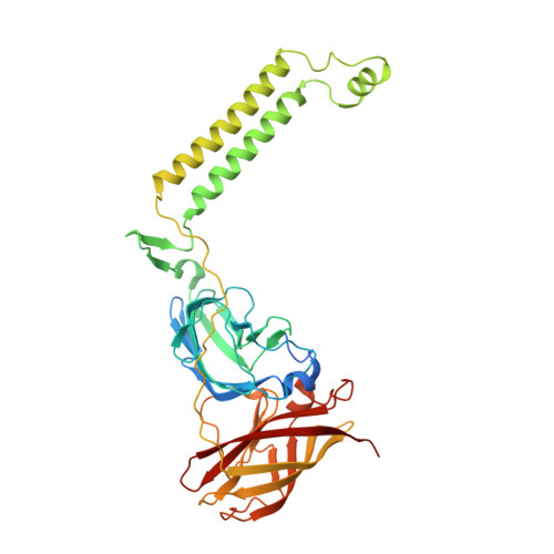
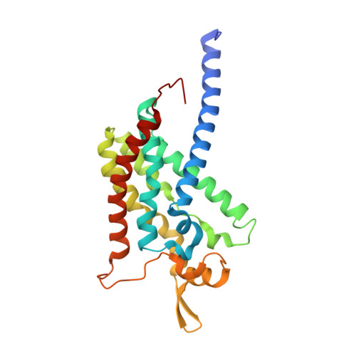
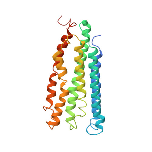
Product analog binding identifies the copper active site of particulate methane monooxygenase.

Tucci, F.J.Jodts, R.J.Hoffman, B.M.Rosenzweig, A.C.

(2023) Nat Catal 6: 1194-1204

  • DOI: https://doi.org/10.1038/s41929-023-01051-x
  • Primary Citation Related Structures: 
    8OYI, 8SQW, 8SR1, 8SR2, 8SR4, 8SR5

  • PubMed Abstract: 

    Nature's primary methane-oxidizing enzyme, the membrane-bound particulate methane monooxygenase (pMMO), catalyzes the oxidation of methane to methanol. pMMO activity requires copper, and decades of structural and spectroscopic studies have sought to identify the active site among three candidates: the Cu B , Cu C , and Cu D sites. Challenges associated with the isolation of active pMMO have hindered progress toward locating its catalytic center. However, reconstituting pMMO into native lipid nanodiscs stabilizes its structure and recovers its activity. Here, these active samples were incubated with 2,2,2,-trifluoroethanol (TFE), a product analog that serves as a readily visualized active-site probe. Interactions of TFE with the Cu D site were observed by both pulsed ENDOR spectroscopy and cryoEM, implicating Cu D and the surrounding hydrophobic pocket as the likely site of methane oxidation. Use of these orthogonal techniques on parallel samples is a powerful approach that can circumvent difficulties in interpreting metalloenzyme cryoEM maps.


  • Organizational Affiliation
    • Departments of Molecular Biosciences and of Chemistry, Northwestern University, Evanston, IL, USA.

Macromolecule Content 

  • Total Structure Weight: 337.22 kDa 
  • Atom Count: 22,882 
  • Modeled Residue Count: 2,577 
  • Deposited Residue Count: 2,763 
  • Unique protein chains: 3

Macromolecules

Find similar proteins by:|  3D Structure
Entity ID: 1
MoleculeChains  Sequence LengthOrganismDetailsImage
Particulate methane monooxygenase alpha subunitA,
D [auth E],
E [auth I]
414Methylococcus capsulatus str. BathMutation(s): 0 
EC: 1.14.18.3
Membrane Entity: Yes 
UniProt
Find proteins for G1UBD1 (Methylococcus capsulatus (strain ATCC 33009 / NCIMB 11132 / Bath))
Explore G1UBD1 
Go to UniProtKB:  G1UBD1
Entity Groups
Sequence Clusters30% Identity50% Identity70% Identity90% Identity95% Identity100% Identity
UniProt GroupG1UBD1
Sequence Annotations
Expand
Reference Sequence
Find similar proteins by:|  3D Structure
Entity ID: 2
MoleculeChains  Sequence LengthOrganismDetailsImage
Particulate methane monooxygenase beta subunitB,
F [auth J],
G [auth F]
247Methylococcus capsulatus str. BathMutation(s): 0 
EC: 1.14.18.3
Membrane Entity: Yes 
UniProt
Find proteins for Q607G3 (Methylococcus capsulatus (strain ATCC 33009 / NCIMB 11132 / Bath))
Explore Q607G3 
Go to UniProtKB:  Q607G3
Entity Groups
Sequence Clusters30% Identity50% Identity70% Identity90% Identity95% Identity100% Identity
UniProt GroupQ607G3
Sequence Annotations
Expand
Reference Sequence
Find similar proteins by:|  3D Structure
Entity ID: 3
MoleculeChains  Sequence LengthOrganismDetailsImage
Ammonia monooxygenase/methane monooxygenase, subunit C family proteinC,
H [auth G],
I [auth K]
260Methylococcus capsulatus str. BathMutation(s): 0 
EC: 1.14.13.25
Membrane Entity: Yes 
UniProt
Find proteins for Q603F1 (Methylococcus capsulatus (strain ATCC 33009 / NCIMB 11132 / Bath))
Explore Q603F1 
Go to UniProtKB:  Q603F1
Entity Groups
Sequence Clusters30% Identity50% Identity70% Identity90% Identity95% Identity100% Identity
UniProt GroupQ603F1
Sequence Annotations
Expand
Reference Sequence

Small Molecules

Ligands 6 Unique
IDChains Name / Formula / InChI Key2D Diagram3D Interactions
PLC

Query on PLC


Download:Ideal Coordinates CCD File
AA [auth C]
AB [auth G]
DA [auth C]
DB [auth G]
GB [auth G]
AA [auth C],
AB [auth G],
DA [auth C],
DB [auth G],
GB [auth G],
HB [auth G],
JB [auth G],
M [auth B],
OB [auth K],
PA [auth J],
QA [auth J],
RB [auth K],
SA [auth F],
SB [auth K],
UA [auth F],
UB [auth K],
V [auth C],
Y [auth C]
DIUNDECYL PHOSPHATIDYL CHOLINE
C32 H65 N O8 P
IJFVSSZAOYLHEE-SSEXGKCCSA-O
P1O

Query on P1O


Download:Ideal Coordinates CCD File
BA [auth C]
CA [auth C]
KA [auth J]
KB [auth G]
LB [auth G]
BA [auth C],
CA [auth C],
KA [auth J],
KB [auth G],
LB [auth G],
N [auth B],
RA [auth J],
S [auth B],
TA [auth F],
VA [auth F],
VB [auth K],
WB [auth K]
1,2-DIDECANOYL-SN-GLYCERO-3-PHOSPHOCHOLINE
C28 H57 N O8 P
MLKLDGSYMHFAOC-AREMUKBSSA-O
HXG

Query on HXG


Download:Ideal Coordinates CCD File
EB [auth G]
IB [auth G]
PB [auth K]
TB [auth K]
W [auth C]
EB [auth G],
IB [auth G],
PB [auth K],
TB [auth K],
W [auth C],
Z [auth C]
1,2-dihexanoyl-sn-glycero-3-phosphocholine
C20 H41 N O8 P
DVZARZBAWHITHR-GOSISDBHSA-O
D10

Query on D10


Download:Ideal Coordinates CCD File
FB [auth G]
GA [auth E]
JA [auth I]
L [auth A]
LA [auth J]
FB [auth G],
GA [auth E],
JA [auth I],
L [auth A],
LA [auth J],
MA [auth J],
NA [auth J],
O [auth B],
OA [auth J],
P [auth B],
Q [auth B],
QB [auth K],
R [auth B],
WA [auth F],
X [auth C],
XA [auth F],
YA [auth F],
ZA [auth F]
DECANE
C10 H22
DIOQZVSQGTUSAI-UHFFFAOYSA-N
CU
(Subject of Investigation/LOI)

Query on CU


Download:Ideal Coordinates CCD File
CB [auth G]
EA [auth E]
FA [auth E]
HA [auth I]
IA [auth I]
CB [auth G],
EA [auth E],
FA [auth E],
HA [auth I],
IA [auth I],
J [auth A],
K [auth A],
NB [auth K],
U [auth C]
COPPER (II) ION
Cu
JPVYNHNXODAKFH-UHFFFAOYSA-N
CL

Query on CL


Download:Ideal Coordinates CCD File
BB [auth G],
MB [auth K],
T [auth C]
CHLORIDE ION
Cl
VEXZGXHMUGYJMC-UHFFFAOYSA-M

Experimental Data & Validation

Experimental Data

  • Method: ELECTRON MICROSCOPY
  • Resolution: 2.36 Å
  • Aggregation State: PARTICLE 
  • Reconstruction Method: SINGLE PARTICLE 

Structure Validation

View Full Validation Report


Entry History 

& Funding Information

Deposition Data


Funding OrganizationLocationGrant Number
National Institutes of Health/National Institute of General Medical Sciences (NIH/NIGMS)United StatesR35GM118035
National Institutes of Health/National Institute of General Medical Sciences (NIH/NIGMS)United StatesR01GM111097
National Institutes of Health/National Institute of General Medical Sciences (NIH/NIGMS)United StatesT32GM105538
National Institutes of Health/National Institute of Environmental Health Sciences (NIH/NIEHS)United StatesF31ES034283
National Institutes of Health/National Institute of General Medical Sciences (NIH/NIGMS)United StatesT32GM008382
National Science Foundation (NSF, United States)United StatesMCB-1908587

Revision History  (Full details and data files)

  • Version 1.0: 2023-11-15
    Type: Initial release
  • Version 2.0: 2024-07-03
    Type: Coordinate replacement
    Reason: Sequence discrepancy
    Changes: Advisory, Atomic model, Author supporting evidence, Data collection, Derived calculations, Source and taxonomy, Structure summary
  • Version 2.1: 2025-03-05
    Changes: Data collection, Database references, Structure summary