8SL7 | pdb_00008sl7

Butyricicoccus sp. BIOML-A1 tryptophanase complex with (3S) ALG-05


Experimental Data Snapshot

  • Method: X-RAY DIFFRACTION
  • Resolution: 2.07 Å
  • R-Value Free: 
    0.247 (Depositor), 0.248 (DCC) 
  • R-Value Work: 
    0.203 (Depositor), 0.204 (DCC) 
  • R-Value Observed: 
    0.204 (Depositor) 

Starting Model: in silico
View more details

wwPDB Validation 3D Report Full Report

Validation slider image for 8SL7

Ligand Structure Quality Assessment 


This is version 1.2 of the entry. See complete history

Literature

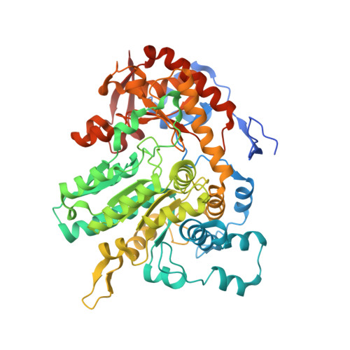
Mechanism-based inhibition of gut microbial tryptophanases reduces serum indoxyl sulfate.

Graboski, A.L.Kowalewski, M.E.Simpson, J.B.Cao, X.Ha, M.Zhang, J.Walton, W.G.Flaherty, D.P.Redinbo, M.R.

(2023) Cell Chem Biol 30: 1402-1413.e7

  • DOI: https://doi.org/10.1016/j.chembiol.2023.07.015
  • Primary Citation Related Structures: 
    8SBG, 8SIJ, 8SL7

  • PubMed Abstract: 

    Indoxyl sulfate is a microbially derived uremic toxin that accumulates in late-stage chronic kidney disease and contributes to both renal and cardiovascular toxicity. Indoxyl sulfate is generated by the metabolism of indole, a compound created solely by gut microbial tryptophanases. Here, we characterize the landscape of tryptophanase enzymes in the human gut microbiome and find remarkable structural and functional similarities across diverse taxa. We leverage this homology through a medicinal chemistry campaign to create a potent pan-inhibitor, (3S) ALG-05, and validate its action as a transition-state analog. (3S) ALG-05 successfully reduces indole production in microbial culture and displays minimal toxicity against microbial and mammalian cells. Mice treated with (3S) ALG-05 show reduced cecal indole and serum indoxyl sulfate levels with minimal changes in other tryptophan-metabolizing pathways. These studies present a non-bactericidal pan-inhibitor of gut microbial tryptophanases with potential promise for reducing indoxyl sulfate in chronic kidney disease.


  • Organizational Affiliation
    • Department of Pharmacology, University of North Carolina, Chapel Hill, Chapel Hill, NC 27599, USA.

Macromolecule Content 

  • Total Structure Weight: 260.64 kDa 
  • Atom Count: 17,306 
  • Modeled Residue Count: 2,171 
  • Deposited Residue Count: 2,276 
  • Unique protein chains: 1

Macromolecules

Find similar proteins by:|  3D Structure
Entity ID: 1
MoleculeChains  Sequence LengthOrganismDetailsImage
Tryptophanase
A, B, C, D
569Butyricicoccus sp. BIOML-A1Mutation(s): 0 
Gene Names: GT642_04165

Experimental Data & Validation

Experimental Data

  • Method: X-RAY DIFFRACTION
  • Resolution: 2.07 Å
  • R-Value Free:  0.247 (Depositor), 0.248 (DCC) 
  • R-Value Work:  0.203 (Depositor), 0.204 (DCC) 
  • R-Value Observed: 0.204 (Depositor) 
Space Group: P 1
Unit Cell:
Length ( Å )Angle ( ˚ )
a = 62.64α = 65.24
b = 93.951β = 88.01
c = 105.604γ = 74.3
Software Package:
Software NamePurpose
PHENIXrefinement
XDSdata reduction
XDSdata scaling
PHASERphasing

Structure Validation

View Full Validation Report


Ligand Structure Quality Assessment 


Entry History 

& Funding Information

Deposition Data


Funding OrganizationLocationGrant Number
National Institutes of Health/National Institute of General Medical Sciences (NIH/NIGMS)United StatesGM135218
National Institutes of Health/National Institute of General Medical Sciences (NIH/NIGMS)United StatesGM137286

Revision History  (Full details and data files)

  • Version 1.0: 2023-08-23
    Type: Initial release
  • Version 1.1: 2023-09-06
    Changes: Database references
  • Version 1.2: 2023-11-29
    Changes: Database references