8SJD

Cryo-EM structure of the Hermes transposase bound to two right-ends of its DNA transposon.

  • Classification: RECOMBINATION/DNA
  • Organism(s): Musca domestica
  • Expression System: Escherichia coli
  • Mutation(s): Yes 

  • Deposited: 2023-04-17 Released: 2023-08-02 
  • Deposition Author(s): Lannes, L., Dyda, F.
  • Funding Organization(s): National Institutes of Health/National Institute of Diabetes and Digestive and Kidney Disease (NIH/NIDDK)

Experimental Data Snapshot

  • Method: ELECTRON MICROSCOPY
  • Resolution: 5.10 Å
  • Aggregation State: PARTICLE 
  • Reconstruction Method: SINGLE PARTICLE 

wwPDB Validation   3D Report Full Report


This is version 1.0 of the entry. See complete history


Literature

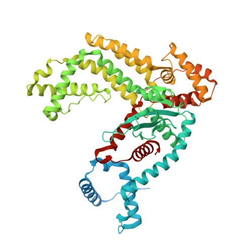
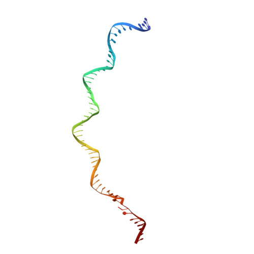
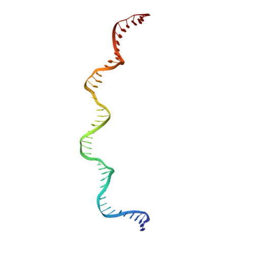
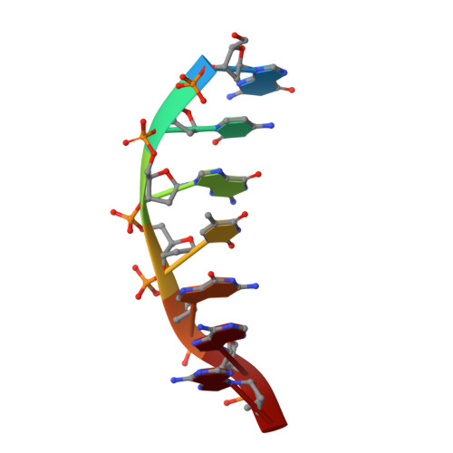
Zinc-finger BED domains drive the formation of the active Hermes transpososome by asymmetric DNA binding.

Lannes, L.Furman, C.M.Hickman, A.B.Dyda, F.

(2023) Nat Commun 14: 4470-4470

  • DOI: https://doi.org/10.1038/s41467-023-40210-3
  • Primary Citation of Related Structures:  
    8EB5, 8EDG, 8SJD

  • PubMed Abstract: 

    The Hermes DNA transposon is a member of the eukaryotic hAT superfamily, and its transposase forms a ring-shaped tetramer of dimers. Our investigation, combining biochemical, crystallography and cryo-electron microscopy, and in-cell assays, shows that the full-length Hermes octamer extensively interacts with its transposon left-end through multiple BED domains of three Hermes protomers contributed by three dimers explaining the role of the unusual higher-order assembly. By contrast, the right-end is bound to no BED domains at all. Thus, this work supports a model in which Hermes multimerizes to gather enough BED domains to find its left-end among the abundant genomic DNA, facilitating the subsequent interaction with the right-end.


  • Organizational Affiliation

    Laboratory of Molecular Biology, National Institute of Diabetes and Digestive and Kidney Diseases, National Institutes of Health, Bethesda, MD, 20892, USA.


Macromolecules

Find similar proteins by:  (by identity cutoff)  |  3D Structure
Entity ID: 1
MoleculeChains Sequence LengthOrganismDetailsImage
Hermes transposaseA [auth C],
B [auth D],
C [auth B],
D [auth A]
612Musca domesticaMutation(s): 2 
UniProt
Find proteins for Q25438 (Musca domestica)
Explore Q25438 
Go to UniProtKB:  Q25438
Entity Groups  
Sequence Clusters30% Identity50% Identity70% Identity90% Identity95% Identity100% Identity
UniProt GroupQ25438
Sequence Annotations
Expand
  • Reference Sequence
Find similar nucleic acids by:  (by identity cutoff)  |  3D Structure
Entity ID: 2
MoleculeChains LengthOrganismImage
DNA (55-MER)E [auth J],
G
55Musca domestica
Sequence Annotations
Expand
  • Reference Sequence
Find similar nucleic acids by:  (by identity cutoff)  |  3D Structure
Entity ID: 3
MoleculeChains LengthOrganismImage
DNA (46-MER)F [auth I],
J [auth F]
46Musca domestica
Sequence Annotations
Expand
  • Reference Sequence

Find similar nucleic acids by:  Sequence   |   3D Structure  

Entity ID: 4
MoleculeChains LengthOrganismImage
DNA (8-MER)H,
I [auth E]
7Musca domestica
Sequence Annotations
Expand
  • Reference Sequence
Experimental Data & Validation

Experimental Data

  • Method: ELECTRON MICROSCOPY
  • Resolution: 5.10 Å
  • Aggregation State: PARTICLE 
  • Reconstruction Method: SINGLE PARTICLE 
EM Software:
TaskSoftware PackageVersion
MODEL REFINEMENTPHENIX1.19.2
RECONSTRUCTIONRELION3.1

Structure Validation

View Full Validation Report


Entry History & Funding Information

Deposition Data


Funding OrganizationLocationGrant Number
National Institutes of Health/National Institute of Diabetes and Digestive and Kidney Disease (NIH/NIDDK)United StatesDK036153-16

Revision History  (Full details and data files)

  • Version 1.0: 2023-08-02
    Type: Initial release