


Crystal structure of PRMT3 with YD1-66
Song, X., Dong, A., Arrowsmith, C.H., Edwards, A.M., Deng, Y., Huang, R., Min, J.To be published.
Experimental Data Snapshot
Starting Model: experimental
View more details
Entity ID: 1 | |||||
|---|---|---|---|---|---|
| Molecule | Chains | Sequence Length | Organism | Details | Image |
| Protein arginine N-methyltransferase 3 | 340 | Homo sapiens | Mutation(s): 0 Gene Names: PRMT3, HRMT1L3 EC: 2.1.1.319 | 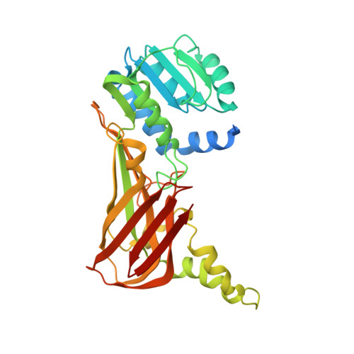 | |
UniProt & NIH Common Fund Data Resources | |||||
PHAROS: O60678 GTEx: ENSG00000185238 | |||||
Entity Groups | |||||
| Sequence Clusters | 30% Identity50% Identity70% Identity90% Identity95% Identity100% Identity | ||||
| UniProt Group | O60678 | ||||
Sequence AnnotationsExpand | |||||
Reference Sequence | |||||
| Ligands 1 Unique | |||||
|---|---|---|---|---|---|
| ID | Chains | Name / Formula / InChI Key | 2D Diagram | 3D Interactions | |
| GXF (Subject of Investigation/LOI) Download:Ideal Coordinates CCD File | C [auth A], D [auth B] | 5'-S-{3-[N'-(4'-chloro[1,1'-biphenyl]-3-yl)carbamimidamido]propyl}-5'-thioadenosine C26 H29 Cl N8 O3 S CEYIEWVKCKYWPH-PTGPVQHPSA-N |  | ||
| Length ( Å ) | Angle ( ˚ ) |
|---|---|
| a = 97.039 | α = 90 |
| b = 100.782 | β = 90 |
| c = 173.437 | γ = 90 |
| Software Name | Purpose |
|---|---|
| PHASER | phasing |
| REFMAC | refinement |
| HKL-3000 | data reduction |
| HKL-3000 | data scaling |
| Funding Organization | Location | Grant Number |
|---|---|---|
| Other private | -- |


