8SGW

Pendrin in complex with chloride

  • Classification: TRANSPORT PROTEIN
  • Organism(s): Sus scrofa
  • Expression System: Trichoplusia ni
  • Mutation(s): No 
  • Membrane Protein: Yes  PDBTMMemProtMD

  • Deposited: 2023-04-13 Released: 2024-02-07 
  • Deposition Author(s): Wang, L., Hoang, A., Zhou, M.
  • Funding Organization(s): National Institutes of Health/National Institute of Diabetes and Digestive and Kidney Disease (NIH/NIDDK), National Institutes of Health/National Institute of General Medical Sciences (NIH/NIGMS)

Experimental Data Snapshot

  • Method: ELECTRON MICROSCOPY
  • Resolution: 2.50 Å
  • Aggregation State: PARTICLE 
  • Reconstruction Method: SINGLE PARTICLE 

wwPDB Validation   3D Report Full Report


This is version 1.1 of the entry. See complete history


Literature

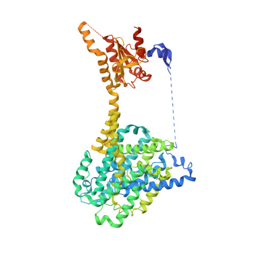
Mechanism of anion exchange and small-molecule inhibition of pendrin.

Wang, L.Hoang, A.Gil-Iturbe, E.Laganowsky, A.Quick, M.Zhou, M.

(2024) Nat Commun 15: 346-346

  • DOI: https://doi.org/10.1038/s41467-023-44612-1
  • Primary Citation of Related Structures:  
    8SGW, 8SH3, 8SHC, 8SIE, 8UUK

  • PubMed Abstract: 

    Pendrin (SLC26A4) is an anion exchanger that mediates bicarbonate (HCO 3 - ) exchange for chloride (Cl - ) and is crucial for maintaining pH and salt homeostasis in the kidney, lung, and cochlea. Pendrin also exports iodide (I - ) in the thyroid gland. Pendrin mutations in humans lead to Pendred syndrome, causing hearing loss and goiter. Inhibition of pendrin is a validated approach for attenuating airway hyperresponsiveness in asthma and for treating hypertension. However, the mechanism of anion exchange and its inhibition by drugs remains poorly understood. We applied cryo-electron microscopy to determine structures of pendrin from Sus scrofa in the presence of either Cl - , I - , HCO 3 - or in the apo-state. The structures reveal two anion-binding sites in each protomer, and functional analyses show both sites are involved in anion exchange. The structures also show interactions between the Sulfate Transporter and Anti-Sigma factor antagonist (STAS) and transmembrane domains, and mutational studies suggest a regulatory role. We also determine the structure of pendrin in a complex with niflumic acid (NFA), which uncovers a mechanism of inhibition by competing with anion binding and impeding the structural changes necessary for anion exchange. These results reveal directions for understanding the mechanisms of anion selectivity and exchange and their regulations by the STAS domain. This work also establishes a foundation for analyzing the pathophysiology of mutations associated with Pendred syndrome.


  • Organizational Affiliation

    Verna and Marrs McLean Department of Biochemistry and Molecular Pharmacology, Baylor College of Medicine, Houston, TX, USA.


Macromolecules
Find similar proteins by:  (by identity cutoff)  |  3D Structure
Entity ID: 1
MoleculeChains Sequence LengthOrganismDetailsImage
PendrinA [auth C],
B [auth D]
780Sus scrofaMutation(s): 0 
Membrane Entity: Yes 
UniProt
Find proteins for A0A8D0Z6H8 (Sus scrofa)
Explore A0A8D0Z6H8 
Go to UniProtKB:  A0A8D0Z6H8
Entity Groups  
Sequence Clusters30% Identity50% Identity70% Identity90% Identity95% Identity100% Identity
UniProt GroupA0A8D0Z6H8
Sequence Annotations
Expand
  • Reference Sequence
Small Molecules
Ligands 5 Unique
IDChains Name / Formula / InChI Key2D Diagram3D Interactions
AV0
Query on AV0

Download Ideal Coordinates CCD File 
NA [auth D],
U [auth C]
Lauryl Maltose Neopentyl Glycol
C47 H88 O22
MADJBYLAYPCCOO-VWHTXWAPSA-N
LBN
Query on LBN

Download Ideal Coordinates CCD File 
AA [auth D]
BA [auth D]
DA [auth D]
EA [auth D]
FA [auth D]
AA [auth D],
BA [auth D],
DA [auth D],
EA [auth D],
FA [auth D],
GA [auth D],
H [auth C],
HA [auth D],
I [auth C],
IA [auth D],
JA [auth D],
K [auth C],
L [auth C],
M [auth C],
N [auth C],
O [auth C],
P [auth C],
Q [auth C]
1-palmitoyl-2-oleoyl-sn-glycero-3-phosphocholine
C42 H82 N O8 P
WTJKGGKOPKCXLL-VYOBOKEXSA-N
DMU
Query on DMU

Download Ideal Coordinates CCD File 
MA [auth D],
T [auth C]
DECYL-BETA-D-MALTOPYRANOSIDE
C22 H42 O11
WOQQAWHSKSSAGF-WXFJLFHKSA-N
CLR
Query on CLR

Download Ideal Coordinates CCD File 
C
CA [auth D]
D [auth C]
E [auth C]
F [auth C]
C,
CA [auth D],
D [auth C],
E [auth C],
F [auth C],
G [auth C],
J [auth C],
V [auth D],
W [auth D],
X [auth D],
Y [auth D],
Z [auth D]
CHOLESTEROL
C27 H46 O
HVYWMOMLDIMFJA-DPAQBDIFSA-N
CL (Subject of Investigation/LOI)
Query on CL

Download Ideal Coordinates CCD File 
KA [auth D],
LA [auth D],
R [auth C],
S [auth C]
CHLORIDE ION
Cl
VEXZGXHMUGYJMC-UHFFFAOYSA-M
Experimental Data & Validation

Experimental Data

  • Method: ELECTRON MICROSCOPY
  • Resolution: 2.50 Å
  • Aggregation State: PARTICLE 
  • Reconstruction Method: SINGLE PARTICLE 

Structure Validation

View Full Validation Report


Entry History & Funding Information

Deposition Data


Funding OrganizationLocationGrant Number
National Institutes of Health/National Institute of Diabetes and Digestive and Kidney Disease (NIH/NIDDK)United StatesDK122784
National Institutes of Health/National Institute of General Medical Sciences (NIH/NIGMS)United StatesGM145416

Revision History  (Full details and data files)

  • Version 1.0: 2024-02-07
    Type: Initial release
  • Version 1.1: 2024-02-28
    Changes: Database references