8SG7

Adeno-Associated Virus Bat origin capsid protein basic regions in complex with importin-alpha 2


Experimental Data Snapshot

  • Method: X-RAY DIFFRACTION
  • Resolution: 2.40 Å
  • R-Value Free: 0.223 
  • R-Value Work: 0.184 
  • R-Value Observed: 0.186 

wwPDB Validation   3D Report Full Report


This is version 1.3 of the entry. See complete history


Literature

Structural basis for nuclear import of bat adeno-associated virus capsid protein.

Hoad, M.Roby, J.A.Forwood, J.K.

(2024) J Gen Virol 105

  • DOI: https://doi.org/10.1099/jgv.0.001960
  • Primary Citation of Related Structures:  
    8SG7

  • PubMed Abstract: 

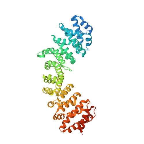
    Adeno-associated viruses (AAV) are one of the world's most promising gene therapy vectors and as a result, are one of the most intensively studied viral vectors. Despite a wealth of research into these vectors, the precise characterisation of AAVs to translocate into the host cell nucleus remains unclear. Recently we identified the nuclear localization signals of an AAV porcine strain and determined its mechanism of binding to host importin proteins. To expand our understanding of diverse AAV import mechanisms we sought to determine the mechanism in which the Cap protein from a bat-infecting AAV can interact with transport receptor importins for translocation into the nucleus. Using a high-resolution crystal structure and quantitative assays, we were able to not only determine the exact region and residues of the N-terminal domain of the Cap protein which constitute the functional NLS for binding with the importin alpha two protein, but also reveal the differences in binding affinity across the importin-alpha isoforms. Collectively our results allow for a detailed molecular view of the way AAV Cap proteins interact with host proteins for localization into the cell nucleus.


  • Organizational Affiliation

    School of Dentistry and Medical Sciences, Charles Sturt University, Wagga Wagga, NSW, 2678, Australia.


Macromolecules
Find similar proteins by:  (by identity cutoff)  |  3D Structure
Entity ID: 1
MoleculeChains Sequence LengthOrganismDetailsImage
Importin subunit alpha-1A [auth B]510Mus musculusMutation(s): 0 
Gene Names: Kpna2Rch1
UniProt & NIH Common Fund Data Resources
Find proteins for P52293 (Mus musculus)
Explore P52293 
Go to UniProtKB:  P52293
IMPC:  MGI:103561
Entity Groups  
Sequence Clusters30% Identity50% Identity70% Identity90% Identity95% Identity100% Identity
UniProt GroupP52293
Sequence Annotations
Expand
  • Reference Sequence
Find similar proteins by:  (by identity cutoff)  |  3D Structure
Entity ID: 2
MoleculeChains Sequence LengthOrganismDetailsImage
VP1 capsidB [auth C]58Bat adeno-associated virusMutation(s): 0 
UniProt
Find proteins for A0A2Z4K5C4 (Bat adeno-associated virus)
Explore A0A2Z4K5C4 
Go to UniProtKB:  A0A2Z4K5C4
Entity Groups  
Sequence Clusters30% Identity50% Identity70% Identity90% Identity95% Identity100% Identity
UniProt GroupA0A2Z4K5C4
Sequence Annotations
Expand
  • Reference Sequence
Experimental Data & Validation

Experimental Data

  • Method: X-RAY DIFFRACTION
  • Resolution: 2.40 Å
  • R-Value Free: 0.223 
  • R-Value Work: 0.184 
  • R-Value Observed: 0.186 
  • Space Group: P 21 21 21
Unit Cell:
Length ( Å )Angle ( ˚ )
a = 79.065α = 90
b = 90.556β = 90
c = 99.816γ = 90
Software Package:
Software NamePurpose
PHENIXrefinement
XDSdata reduction
Aimlessdata scaling
PHASERphasing

Structure Validation

View Full Validation Report


Entry History & Funding Information

Deposition Data


Funding OrganizationLocationGrant Number
Not funded--

Revision History  (Full details and data files)

  • Version 1.0: 2023-07-19
    Type: Initial release
  • Version 1.1: 2023-09-27
    Changes: Data collection
  • Version 1.2: 2024-01-10
    Changes: Data collection, Experimental preparation
  • Version 1.3: 2024-03-20
    Changes: Database references