8SDB

Crystal Structure of E.Coli Branching Enzyme in complex with malto-octose


Experimental Data Snapshot

  • Method: X-RAY DIFFRACTION
  • Resolution: 3.00 Å
  • R-Value Free: 0.315 
  • R-Value Work: 0.214 
  • R-Value Observed: 0.219 

wwPDB Validation   3D Report Full Report


Ligand Structure Quality Assessment 


This is version 1.1 of the entry. See complete history


Literature

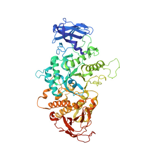
The Structure of Maltooctaose-Bound Escherichia coli Branching Enzyme Suggests a Mechanism for Donor Chain Specificity.

Fawaz, R.Bingham, C.Nayebi, H.Chiou, J.Gilbert, L.Park, S.H.Geiger, J.H.

(2023) Molecules 28

  • DOI: https://doi.org/10.3390/molecules28114377
  • Primary Citation of Related Structures:  
    8SDB

  • PubMed Abstract: 

    Glycogen is the primary storage polysaccharide in bacteria and animals. It is a glucose polymer linked by α-1,4 glucose linkages and branched via α-1,6-linkages, with the latter reaction catalyzed by branching enzymes. Both the length and dispensation of these branches are critical in defining the structure, density, and relative bioavailability of the storage polysaccharide. Key to this is the specificity of branching enzymes because they define branch length. Herein, we report the crystal structure of the maltooctaose-bound branching enzyme from the enterobacteria E. coli . The structure identifies three new malto-oligosaccharide binding sites and confirms oligosaccharide binding in seven others, bringing the total number of oligosaccharide binding sites to twelve. In addition, the structure shows distinctly different binding in previously identified site I, with a substantially longer glucan chain ordered in the binding site. Using the donor oligosaccharide chain-bound Cyanothece branching enzyme structure as a guide, binding site I was identified as the likely binding surface for the extended donor chains that the E. coli branching enzyme is known to transfer. Furthermore, the structure suggests that analogous loops in branching enzymes from a diversity of organisms are responsible for branch chain length specificity. Together, these results suggest a possible mechanism for transfer chain specificity involving some of these surface binding sites.


  • Organizational Affiliation

    Department of Chemistry, Michigan State University, East Lansing, MI 48824, USA.


Macromolecules
Find similar proteins by:  (by identity cutoff)  |  3D Structure
Entity ID: 1
MoleculeChains Sequence LengthOrganismDetailsImage
1,4-alpha-glucan branching enzyme GlgB
A, B, C, D
613Escherichia coliMutation(s): 0 
Gene Names: glgBb3432JW3395
EC: 2.4.1.18
UniProt
Find proteins for P07762 (Escherichia coli (strain K12))
Explore P07762 
Go to UniProtKB:  P07762
Entity Groups  
Sequence Clusters30% Identity50% Identity70% Identity90% Identity95% Identity100% Identity
UniProt GroupP07762
Sequence Annotations
Expand
  • Reference Sequence
Oligosaccharides

Help

Entity ID: 2
MoleculeChains Length2D Diagram Glycosylation3D Interactions
alpha-D-glucopyranose-(1-4)-alpha-D-glucopyranose-(1-4)-alpha-D-glucopyranose-(1-4)-alpha-D-glucopyranose-(1-4)-alpha-D-glucopyranose
E, F, H
5N/A
Glycosylation Resources
GlyTouCan:  G50146AM
GlyCosmos:  G50146AM
Entity ID: 3
MoleculeChains Length2D Diagram Glycosylation3D Interactions
alpha-D-glucopyranose-(1-4)-alpha-D-glucopyranose-(1-4)-alpha-D-glucopyranose-(1-4)-alpha-D-glucopyranose
G
4N/A
Glycosylation Resources
GlyTouCan:  G87171PZ
GlyCosmos:  G87171PZ
GlyGen:  G87171PZ
Entity ID: 4
MoleculeChains Length2D Diagram Glycosylation3D Interactions
alpha-D-glucopyranose-(1-4)-alpha-D-glucopyranose-(1-4)-alpha-D-glucopyranose
I, M
3N/A
Glycosylation Resources
GlyTouCan:  G96370VA
GlyCosmos:  G96370VA
GlyGen:  G96370VA
Entity ID: 5
MoleculeChains Length2D Diagram Glycosylation3D Interactions
alpha-D-glucopyranose-(1-4)-alpha-D-glucopyranose-(1-4)-alpha-D-glucopyranose-(1-4)-alpha-D-glucopyranose-(1-4)-alpha-D-glucopyranose-(1-4)-alpha-D-glucopyranose-(1-4)-alpha-D-glucopyranose
J
7N/A
Glycosylation Resources
GlyTouCan:  G41288IQ
GlyCosmos:  G41288IQ
GlyGen:  G41288IQ
Entity ID: 6
MoleculeChains Length2D Diagram Glycosylation3D Interactions
alpha-D-glucopyranose-(1-4)-alpha-D-glucopyranose
K, L, N
2N/A
Glycosylation Resources
GlyTouCan:  G07411ON
GlyCosmos:  G07411ON
Experimental Data & Validation

Experimental Data

  • Method: X-RAY DIFFRACTION
  • Resolution: 3.00 Å
  • R-Value Free: 0.315 
  • R-Value Work: 0.214 
  • R-Value Observed: 0.219 
  • Space Group: P 32 2 1
Unit Cell:
Length ( Å )Angle ( ˚ )
a = 146.489α = 90
b = 146.489β = 90
c = 294.92γ = 120
Software Package:
Software NamePurpose
Cootmodel building
PHENIXrefinement
HKL-2000data reduction
HKL-2000data scaling
PHENIXphasing

Structure Validation

View Full Validation Report


Ligand Structure Quality Assessment 


Entry History & Funding Information

Deposition Data


Funding OrganizationLocationGrant Number
National Institutes of Health/National Institute of Biomedical Imaging and Bioengineering (NIH/NIBIB)United States--
Department of Energy (DOE, United States)United States--

Revision History  (Full details and data files)

  • Version 1.0: 2023-07-12
    Type: Initial release
  • Version 1.1: 2023-08-16
    Changes: Data collection