8SCA | pdb_00008sca

Rec3 Domain from S. pyogenes Cas9


Experimental Data Snapshot

  • Method: X-RAY DIFFRACTION
  • Resolution: 1.67 Å
  • R-Value Free: 
    0.209 (Depositor), 0.209 (DCC) 
  • R-Value Work: 
    0.186 (Depositor), 0.186 (DCC) 
  • R-Value Observed: 
    0.187 (Depositor) 

Starting Model: experimental
View more details

wwPDB Validation 3D Report Full Report

Validation slider image for 8SCA

This is version 1.0 of the entry. See complete history

Literature

High-fidelity, hyper-accurate, and evolved mutants rewire atomic-level communication in CRISPR-Cas9.

Skeens, E.Sinha, S.Ahsan, M.D'Ordine, A.M.Jogl, G.Palermo, G.Lisi, G.P.

(2024) Sci Adv 10: eadl1045-eadl1045

  • DOI: https://doi.org/10.1126/sciadv.adl1045
  • Primary Citation Related Structures: 
    8SCA

  • PubMed Abstract: 

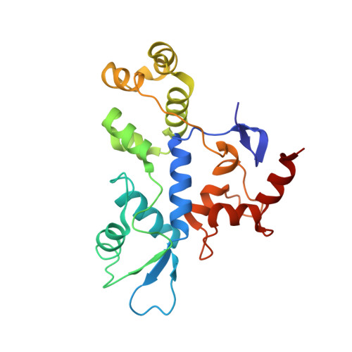
    The high-fidelity (HF1), hyper-accurate (Hypa), and evolved (Evo) variants of the CRISPR-associated protein 9 (Cas9) endonuclease are critical tools to mitigate off-target effects in the application of CRISPR-Cas9 technology. The mechanisms by which mutations in recognition subdomain 3 (Rec3) mediate specificity in these variants are poorly understood. Here, solution nuclear magnetic resonance and molecular dynamics simulations establish the structural and dynamic effects of high-specificity mutations in Rec3, and how they propagate the allosteric signal of Cas9. We reveal conserved structural changes and dynamic differences at regions of Rec3 that interface with the RNA:DNA hybrid, transducing chemical signals from Rec3 to the catalytic His-Asn-His (HNH) domain. The variants remodel the communication sourcing from the Rec3 α helix 37, previously shown to sense target DNA complementarity, either directly or allosterically. This mechanism increases communication between the DNA mismatch recognition helix and the HNH active site, shedding light on the structure and dynamics underlying Cas9 specificity and providing insight for future engineering principles.


  • Organizational Affiliation
    • Department of Molecular Biology, Cell Biology and Biochemistry, Brown University, Providence, RI, USA.

Macromolecule Content 

  • Total Structure Weight: 26.13 kDa 
  • Atom Count: 2,033 
  • Modeled Residue Count: 218 
  • Deposited Residue Count: 218 
  • Unique protein chains: 1

Macromolecules

Find similar proteins by:|  3D Structure
Entity ID: 1
MoleculeChains  Sequence LengthOrganismDetailsImage
CRISPR-associated endonuclease Cas9/Csn1A [auth B]218Streptococcus pyogenesMutation(s): 0 
Gene Names: cas9csn1SPy_1046
EC: 3.1
UniProt
Find proteins for Q99ZW2 (Streptococcus pyogenes serotype M1)
Explore Q99ZW2 
Go to UniProtKB:  Q99ZW2
Entity Groups
Sequence Clusters30% Identity50% Identity70% Identity90% Identity95% Identity100% Identity
UniProt GroupQ99ZW2
Sequence Annotations
Expand
Reference Sequence

Experimental Data & Validation

Experimental Data

  • Method: X-RAY DIFFRACTION
  • Resolution: 1.67 Å
  • R-Value Free:  0.209 (Depositor), 0.209 (DCC) 
  • R-Value Work:  0.186 (Depositor), 0.186 (DCC) 
  • R-Value Observed: 0.187 (Depositor) 
Space Group: P 21 21 21
Unit Cell:
Length ( Å )Angle ( ˚ )
a = 53.36α = 90
b = 54.631β = 90
c = 73.8γ = 90
Software Package:
Software NamePurpose
XDSdata reduction
Aimlessdata scaling
PHASERphasing
PHENIXrefinement

Structure Validation

View Full Validation Report


Entry History 

& Funding Information

Deposition Data


Funding OrganizationLocationGrant Number
National Institutes of Health/National Institute of General Medical Sciences (NIH/NIGMS)United States--

Revision History  (Full details and data files)

  • Version 1.0: 2024-03-13
    Type: Initial release