


Structural basis of Arf1-driven membrane tubulation
Haupt, C., Semchonok, D.A., Desfosses, A., Daum, S., Neudorf, S.S., Hamdi, F., Kastritis, P.L., Stubbs, M.T., Bacia, K.(2026) bioRxiv
Experimental Data Snapshot
Starting Model: experimental
View more details
wwPDB Validation 3D Report Full Report

(2026) bioRxiv
Entity ID: 1 | |||||
|---|---|---|---|---|---|
| Molecule | Chains | Sequence Length | Organism | Details | Image |
| ADP-ribosylation factor 1 | 181 | Saccharomyces cerevisiae | Mutation(s): 0 Gene Names: ARF1, YDL192W, D1244 EC: 3.6.5.2 | 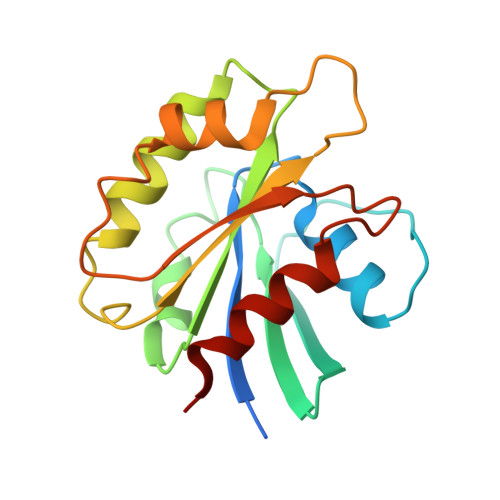 | |
UniProt | |||||
Entity Groups | |||||
| Sequence Clusters | 30% Identity50% Identity70% Identity90% Identity95% Identity100% Identity | ||||
| UniProt Group | P11076 | ||||
Sequence AnnotationsExpand | |||||
Reference Sequence | |||||
| Ligands 2 Unique | |||||
|---|---|---|---|---|---|
| ID | Chains | Name / Formula / InChI Key | 2D Diagram | 3D Interactions | |
| GSP (Subject of Investigation/LOI) Download:Ideal Coordinates CCD File | I [auth A] K [auth B] M [auth C] O [auth D] Q [auth E] | 5'-GUANOSINE-DIPHOSPHATE-MONOTHIOPHOSPHATE C10 H16 N5 O13 P3 S XOFLBQFBSOEHOG-UUOKFMHZSA-N |  | ||
| MG (Subject of Investigation/LOI) Download:Ideal Coordinates CCD File | H [auth A] J [auth B] L [auth C] N [auth D] P [auth E] | MAGNESIUM ION Mg JLVVSXFLKOJNIY-UHFFFAOYSA-N |  | ||
| Task | Software Package | Version |
|---|---|---|
| RECONSTRUCTION | cryoSPARC | v4.3.1 |
| MODEL REFINEMENT | PHENIX | 1.21_5187 |
| Funding Organization | Location | Grant Number |
|---|---|---|
| German Federal Ministry for Education and Research | Germany | 03Z22HN23 |
| German Federal Ministry for Education and Research | Germany | 03Z22HI2 |


