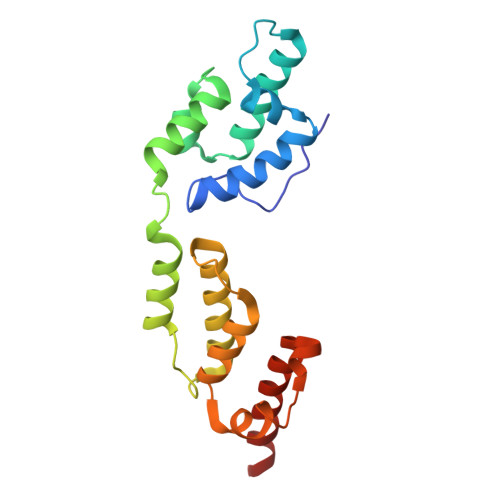
In-cell structure and snapshots of copia retrotransposons in intact tissue by cryo-ET.
Klumpe, S., Senti, K.A., Beck, F., Sachweh, J., Hampoelz, B., Ronchi, P., Oorschot, V., Brandstetter, M., Yeroslaviz, A., Briggs, J.A.G., Brennecke, J., Beck, M., Plitzko, J.M.(2025) Cell 188: 2094
- PubMed: 40049165 Search on PubMed
- DOI: https://doi.org/10.1016/j.cell.2025.02.003
- Primary Citation Related Structures:
8S41 - PubMed Abstract:
Long terminal repeat (LTR) retrotransposons belong to the transposable elements (TEs), autonomously replicating genetic elements that integrate into the host's genome. Among animals, Drosophila melanogaster serves as an important model organism for TE research and contains several LTR retrotransposons, including the Ty1-copia family, which is evolutionarily related to retroviruses and forms virus-like particles (VLPs). In this study, we use cryo-focused ion beam (FIB) milling and lift-out approaches to visualize copia VLPs in ovarian cells and intact egg chambers, resolving the in situ copia capsid structure to 7.7 Å resolution by cryoelectron tomography (cryo-ET). Although cytoplasmic copia VLPs vary in size, nuclear VLPs are homogeneous and form densely packed clusters, supporting a model in which nuclear import acts as a size selector. Analyzing flies deficient in the TE-suppressing PIWI-interacting RNA (piRNA) pathway, we observe copia's translocation into the nucleus during spermatogenesis. Our findings provide insights into the replication cycle and cellular structural biology of an active LTR retrotransposon.
- Research Group CryoEM Technology, Max Planck Institute of Biochemistry, Martinsried, Germany. Electronic address: klumpe@biochem.mpg.de.
Organizational Affiliation: