8S1K | pdb_00008s1k

Crystal Structure of human FABP4 in complex with 2-[1-(methoxymethyl)cyclopentyl]-6-pentyl-4-phenyl-3-(1H-tetrazol-5-yl)-5,6,7,8-tetrahydroquinoline


Experimental Data Snapshot

  • Method: X-RAY DIFFRACTION
  • Resolution: 1.22 Å
  • R-Value Free: 
    0.207 (Depositor), 0.210 (DCC) 
  • R-Value Work: 
    0.171 (Depositor), 0.190 (DCC) 
  • R-Value Observed: 
    0.173 (Depositor) 

wwPDB Validation   3D Report Full Report


Ligand Structure Quality Assessment 


This is version 1.1 of the entry. See complete history


Literature

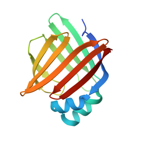
A high-resolution data set of fatty acid-binding protein structures. I. Dynamics of FABP4 and ligand binding.

Casagrande, F.Ehler, A.Burger, D.Benz, J.Ross, A.Rudolph, M.G.

(2025) Acta Crystallogr D Struct Biol 81: 423-435

  • DOI: https://doi.org/10.1107/S2059798325006242
  • Primary Citation of Related Structures:  
    8S1K

  • PubMed Abstract: 

    Fatty acid-binding proteins (FABPs) are involved in the uptake and intracellular trafficking of fatty acids for metabolic and gene-regulatory purposes. FABPs are known to associate with membranes and also enter the nucleus. Using NMR and a human FABP4 (hFABP4) preparation completely free of endogenous ligands, we studied the influence of fatty acids and inhibitors on the conformational flexibility and bicelle/membrane association of this isoform. Binding of fatty acids and ligands rigidifies hFABP4, particularly at the portal region where ligands enter the binding site. Depending on the nature of the ligand, hFABP4 stays associated with bicelles via the portal region or segregates into solution, a prerequisite for nuclear import using a nonclassical nuclear localization signal. These results indicate that different ligands can lead to different biological outcomes. One of the major determinants for FABP4 segregation is Phe58, which in X-ray crystal structures adopts different conformations as a function of ligand volume. It is possible that other FABP isoforms use a similar mechanism for ligand-dependent membrane detachment and activation of nuclear import.


  • Organizational Affiliation
    • Therapeutic Modalities, Innovation Center Basel, F. Hoffmann-La Roche, Grenzacherstrasse 124, 4070 Basel, Switzerland.

Macromolecules
Find similar proteins by:  (by identity cutoff)  |  3D Structure
Entity ID: 1
MoleculeChains Sequence LengthOrganismDetailsImage
Fatty acid-binding protein, adipocyte135Homo sapiensMutation(s): 0 
Gene Names: FABP4
UniProt & NIH Common Fund Data Resources
Find proteins for P15090 (Homo sapiens)
Explore P15090 
Go to UniProtKB:  P15090
PHAROS:  P15090
GTEx:  ENSG00000170323 
Entity Groups  
Sequence Clusters30% Identity50% Identity70% Identity90% Identity95% Identity100% Identity
UniProt GroupP15090
Sequence Annotations
Expand
  • Reference Sequence
Small Molecules
Ligands 2 Unique
IDChains Name / Formula / InChI Key2D Diagram3D Interactions
A1H4Y (Subject of Investigation/LOI)
Query on A1H4Y

Download Ideal Coordinates CCD File 
B [auth A](6~{S})-2-[1-(methoxymethyl)cyclopentyl]-6-pentyl-4-phenyl-3-(1~{H}-1,2,3,4-tetrazol-5-yl)-5,6,7,8-tetrahydroquinoline
C28 H37 N5 O
OGYHUAQNUJEKHA-FQEVSTJZSA-N
DMS
Query on DMS

Download Ideal Coordinates CCD File 
C [auth A]DIMETHYL SULFOXIDE
C2 H6 O S
IAZDPXIOMUYVGZ-UHFFFAOYSA-N
Experimental Data & Validation

Experimental Data

  • Method: X-RAY DIFFRACTION
  • Resolution: 1.22 Å
  • R-Value Free:  0.207 (Depositor), 0.210 (DCC) 
  • R-Value Work:  0.171 (Depositor), 0.190 (DCC) 
  • R-Value Observed: 0.173 (Depositor) 
Space Group: P 2 21 21
Unit Cell:
Length ( Å )Angle ( ˚ )
a = 31.988α = 90
b = 53.4β = 90
c = 72.131γ = 90
Software Package:
Software NamePurpose
XSCALEdata scaling
PHENIXrefinement
PDB_EXTRACTdata extraction
XDSdata reduction
PHASERphasing

Structure Validation

View Full Validation Report


Ligand Structure Quality Assessment 


Entry History & Funding Information

Deposition Data


Funding OrganizationLocationGrant Number
F. Hoffmann-La Roche LTDSwitzerland--

Revision History  (Full details and data files)

  • Version 1.0: 2024-03-06
    Type: Initial release
  • Version 1.1: 2025-09-17
    Changes: Database references, Structure summary